Page 245 - Feline Cardiology
P. 245
250  Section F: Arrhythmias and Other Electrocardiographic Abnormalities


                                                                                                      QRS


                                                                                                  QRS
                                                                                                     T
                                                                                                 P      P


                                                                                                          T
              A


                                                                                                  QRS     QRS
                                                                                               T      P  T    P
                                                                                            P

      Arrhythmias


              B
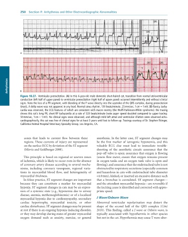
              Figure 18.27.  Ventricular	preexcitation.	(A)	In	this	6-year-old	male	domestic	short-haired	cat,	transition	from	normal	atrioventricular
              conduction	(left	half	of	upper	panel)	to	ventricular	preexcitation	(right	half	of	upper	panel)	occurred	intermittently	and	without	clinical
              signs.	Note	the	loss	of	a	PR	segment,	with	blending	of	the	P	wave	directly	into	the	upstroke	of	the	QRS	complex,	during	preexcitation
              (inset).	A	delta	wave	was	not	apparent	in	any	lead.	Normal	sinus	rhythm,	130	beats/minute.	25	mm/sec,	1	cm	=	1	mV.	(B)	Rarely,	tachy-
              cardia	was	observed,	the	ECG	features	of	which	are	consistent	with	macro	reentry	(the	Wolff-Parkinson-White	syndrome):	the	tracing
              shows	this	cat’s	long	PR,	short	RP	tachycardia	at	a	rate	of	320	beats/minute	(note	paper	speed	doubled	compared	to	upper	tracing:
              50	mm/sec,	1	cm	=	1	mV).	No	clinical	signs	were	observed,	and	although	mild	left	atrial	and	ventricular	dilation	were	observed	echo-
              cardiographically,	this	cat	was	free	of	clinical	signs	for	at	least	3	years	until	lost	to	follow-up.	Tracings	courtesy	of	Dr.	Stephen	Ettinger,
              California	Animal	Hospital	Veterinary	Specialty	Group,	Los	Angeles,	CA.


                 zones  that  leads  to  current  flow  between  these   anesthesia. In the latter case, ST segment changes may
                 regions. These currents of injury are represented   be  the  first  marker  of  iatrogenic  hypoxemia,  and  this
                 on the surface ECG by deviation of the ST segment   valuable  ECG  clue  must  lead  to  immediate  trouble-
                 (Mirvis and Goldberger 2008).                   shooting  of  the  anesthetic  circuit:  assurance  that  the
                                                                 pop-off valve is open; assurance that oxygen is flowing
                 This principle is based on regional or uneven zones   (assess flow meter, ensure that oxygen remains present
              of ischemia, which is likely to occur even in the absence   in oxygen tanks and an oxygen tank valve is open and
              of coronary artery disease according to several mecha-  flowing); and assurance that the endotracheal tube is not
              nisms,  including  coronary  vasospasm,  regional  varia-  obstructed by respiratory secretions (especially common
              tions  in  myocardial  blood  flow,  and  heterogeneity  of   and hazardous in cats with endotracheal tube diameter
              myocardial thickness.                              <4.0 mm), kinked, or inserted an excessive distance such
                 In feline practice, ST segment changes are important   that  a  bronchus  is  cannulated.  ST  segment  changes—
              because  they  can  constitute  a  marker  for  myocardial   and the attendant myocardial hypoxia—are reversible if
              hypoxia. ST segment changes in cats may be an expres-  the inciting cause is identified and corrected with appro-
              sion of a systemic state (e.g., hypoxemia due to airway   priate speed.
              disease, anemia, methemoglobinemia) or of a localized
              myocardial hypoxia due to cardiomyopathy, secondary   J Wave/Osborn Wave
              cardiac  hypertrophy,  myocardial  toxicity,  or  other   Abnormal  ventricular  repolarization  may  distort  the
              cardiac disturbance. ST segment changes may be present   shape  of  the  second  half  of  the  QRS  complex  (Côté
              at rest if there is an ongoing hypoxia-inducing disorder,   2010). This finding, called a J wave or Osborn wave, is
              or they may develop during states of greater myocardial   typically associated with hypothermia in other species
              oxygen  demand  such  as  anxiety,  exercise,  or  general   but not in the cat. Hypothermia may cause T wave alter-

