Page 242 - Feline Cardiology
P. 242
T    T
                                                                                                            R


               A                                                                                            S


                                                                                                        T     T
                                                                                                           R
                                                                                                          P


                                                                                                            S
               B                                                                                                        Arrhythmias


                                                                                                          T   T


               C                                                                                           QS


                                                                                                          R
                                                                                                     T      T
                                                                                                        P
                                                                                                           S


               D
                                                                                                +
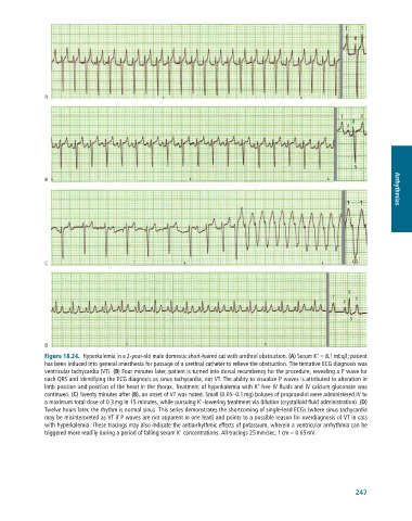
              Figure 18.24.  Hyperkalemia	in	a	2-year-old	male	domestic	short-haired	cat	with	urethral	obstruction.	(A)	Serum	K 	=	8.1	mEq/l;	patient
              has	been	induced	into	general	anesthesia	for	passage	of	a	urethral	catheter	to	relieve	the	obstruction.	The	tentative	ECG	diagnosis	was
              ventricular	tachycardia	(VT).	(B)	Four	minutes	later,	patient	is	turned	into	dorsal	recumbency	for	the	procedure,	revealing	a	P	wave	for
              each	QRS	and	identifying	the	ECG	diagnosis	as	sinus	tachycardia,	not	VT.	The	ability	to	visualize	P	waves	is	attributed	to	alteration	in
                                                                               +
              limb	position	and	position	of	the	heart	in	the	thorax.	Treatment	of	hyperkalemia	with	K -free	IV	fluids	and	IV	calcium	gluconate	was
              continued.	(C)	Twenty	minutes	after	(B),	an	onset	of	VT	was	noted.	Small	(0.05–0.1	mg)	boluses	of	propranolol	were	administered	IV	to
                                                             +
              a	maximum	total	dose	of	0.3	mg	in	15	minutes,	while	pursuing	K -lowering	treatment	via	dilution	(crystalloid	fluid	administration).	(D)
              Twelve	hours	later,	the	rhythm	is	normal	sinus.	This	series	demonstrates	the	shortcoming	of	single-lead	ECGs	(where	sinus	tachycardia
              may	be	misinterpreted	as	VT	if	P	waves	are	not	apparent	in	one	lead)	and	points	to	a	possible	reason	for	overdiagnosis	of	VT	in	cats
              with	hyperkalemia.	These	tracings	may	also	indicate	the	antiarrhythmic	effects	of	potassium,	wherein	a	ventricular	arrhythmia	can	be
                                                       +
              triggered	more	readily	during	a	period	of	falling	serum	K 	concentrations.	All	tracings	25	mm/sec,	1	cm	=	0.65	mV.


                                                                                                            247

