Page 237 - Feline Cardiology
P. 237
242  Section F: Arrhythmias and Other Electrocardiographic Abnormalities


                I             II            III            aVR           aVL           aVF           V2


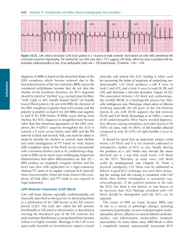
              Figure 18.22.  Left	anterior	fascicular	(LAF)	block	pattern	in	a	13-year-old	male	domestic	short-haired	cat	with	mild	symmetrical	left
              ventricular	concentric	hypertrophy.	The	marked	left	axis	shift	seen	here	(−75°)	suggests	LAF	block,	which	has	been	associated	with	hy-
              pertrophic	cardiomyopathy	in	cats.	Sinus	tachycardia;	heart	rate	=	230	beats/minute.	25	mm/sec,	1	cm	=	1	mV.


              diagnosis of BBBs is based on the abnormal shape of the   clinically,  and  indeed  this  ECG  finding  is  rather  used
              QRS  complexes,  which  become  widened  due  to  the   for increasing the index of suspicion of underlying car-
      Arrhythmias  considered  arrhythmias  because  they  do  not  alter  the   leads I and aVL, and a deep S wave in leads II, III, and
                                                                 diomyopathy.  LAF  block  produces  a  tall  R  wave  in
              desynchronization of the two ventricles. BBBs cannot be
                                                                 aVF, and therefore a left-axis deviation (Figure 18.22).
              rhythm of the heartbeat; therefore, the ECG diagnosis
              should be stated as “rhythm” (e.g., normal sinus rhythm)
                                                                 thy, notably HCM, is a histologically proven but clini-
              “with [right or left] bundle branch block” (or bundle   The association between LAF block and cardiomyopa-
              branch block pattern). In cats with BBB, the duration of   cally ambiguous one. Histologic observation of fibrosis
              the QRS complexes is greater than 0.04 second, and the   involving  especially  the  left  parts  of  the  His-Purkinje
              polarity is positive in lead II for left BBBs and negative   system  in  cats  with  HCM  supports  the  link  between
              in lead II for RBB blocks. If BBBs occur during sinus   HCM and LAF block (Kaneshige et al. 2006); a survey
              rhythm, the ECG diagnosis is straightforward, because   of 63 cardiomyopathic feline hearts revealed degenera-
              other than the abnormal appearance of the QRS com-  tion, fibrosis, osseous metaplasia, and other lesions in 58
              plexes,  the  P-QRS-T  sequence  throughout  the  ECG  is   (92%) of cases, with 54 (86%) of left bundles affected
              normal: a P wave occurs before each QRS and the PR   compared to only 20 (32%) of right bundles (Liu et al.
              interval is fixed and normal. Still, care must be taken to   1975).
              properly  identify  the  rhythm  as  normal  sinus  rhythm   It should be noted that an important artifact could
              and  avoid  misdiagnosis  of VT  based  on  wide,  bizarre   mimic LAF block and it is not routinely addressed in
              QRS complexes alone. If the block occurs concurrently   retrospective  studies  of  ECG  in  cats.  Simply  altering
              with a nonsinus rhythm, such as AF, establishing a diag-  the  position  of  a  cat’s  limbs  may  deviate  the  mean
              nosis in BBB can be much more challenging. Important   electrical  axis  in  a  way  that  could  mimic  LAF  block
              characteristics that allow differentiation are that AF +   on  the  ECG.  Therefore,  in  some  cases,  LAF  block
              BBB  produce  an  irregularly  irregular  rhythm  and  the   could  be  misdiagnosed  (see  Chapter  9).  From  a
              heart may slow with application of a vagal maneuver,   practical  standpoint,  LAF  block  may  be  addressed  as
              whereas VT tends to be regular (constant R-R interval)   follows: if good ECG technique was used when obtain-
              when monomorphic (when the wide, bizarre QRS com-  ing the tracing and the tracing is consistent with LAF
              plexes all look alike) and VT tends to not respond to   block,  then  further  investigation  is  warranted  (e.g.,
              vagal maneuvers.                                   echocardiogram).  If  the  patient’s  position  at  the  time
                                                                 the  ECG  was  done  is  not  known  or  was  known  to
              Left Anterior Fascicular (LAF) Block               be  incorrect,  then  ECG  findings  consistent  with  LAF
              Cats with heart disease, especially cardiomyopathy, are   block  should  be  disregarded  until  the  ECG  can  be
              classically described as being prone to developing block   repeated.
              in a subdivision of the LBB known as the left anterior   The  causes  of  BBB  are  many,  because  BBBs  may
              fascicle  (LAF).  The  LAF,  also  called  the  left  anterior   be  due  to  a  variety  of  pathologic  changes,  including
              hemibundle or left anterior arborization, is described as   concentric hypertrophy (as seen in hypertrophic cardio-
              covering  the  dorsalmost  part  of  the  left  ventricle,  but   myopathy, above), dilation (as seen in dilated cardiomy-
              such anatomic distribution is extrapolated from humans   opathy),  and  inflammation  (endocarditis,  traumatic
              (where it is highly variable). Blockage of the LAF is not   myocarditis). In many feline cases, RBB block is often
              appreciably harmful: no hemodynamic impact is noted   a  completely  normal,  unnecessarily  worrisome  ECG

