Page 217 - Feline Cardiology
P. 217
222  Section F: Arrhythmias and Other Electrocardiographic Abnormalities


                                                                                                          QRS


                                                                                                        P
                                                                                                              T


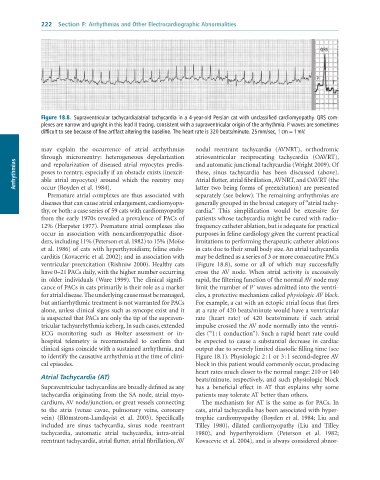
              Figure 18.8.  Supraventricular	tachycardia/atrial	tachycardia	in	a	4-year-old	Persian	cat	with	unclassified	cardiomyopathy.	QRS	com-
              plexes	are	narrow	and	upright	in	this	lead	II	tracing,	consistent	with	a	supraventricular	origin	of	the	arrhythmia.	P	waves	are	sometimes
              difficult	to	see	because	of	fine	artifact	altering	the	baseline.	The	heart	rate	is	320	beats/minute.	25	mm/sec,	1	cm	=	1	mV.


              may  explain  the  occurrence  of  atrial  arrhythmias   nodal  reentrant  tachycardia  (AVNRT),  orthodromic
              through  microreentry:  heterogeneous  depolarization   atrioventricular  reciprocating  tachycardia  (OAVRT),
      Arrhythmias  poses to reentry, especially if an obstacle exists (inexcit-  these,  sinus  tachycardia  has  been  discussed  (above).
              and  repolarization  of  diseased  atrial  myocytes  predis-
                                                                 and automatic junctional tachycardia (Wright 2009). Of
                                                                 Atrial flutter, atrial fibrillation, AVNRT, and OAVRT (the
              able  atrial  myocytes)  around  which  the  reentry  may
              occur (Boyden et al. 1984).
                                                                 separately (see below). The remaining arrhythmias are
                 Premature atrial complexes are thus associated with   latter two being forms of preexcitation) are presented
              diseases that can cause atrial enlargement, cardiomyopa-  generally grouped in the broad category of “atrial tachy-
              thy, or both: a case series of 59 cats with cardiomyopathy   cardia.”  This  simplification  would  be  excessive  for
              from the early 1970s revealed a prevalence of PACs of   patients whose tachycardia might be cured with radio-
              12% (Harpster 1977). Premature atrial complexes also   frequency catheter ablation, but is adequate for practical
              occur  in  association  with  noncardiomyopathic  disor-  purposes in feline cardiology given the current practical
              ders, including 11% (Peterson et al. 1982) to 15% (Moïse   limitations to performing therapeutic catheter ablations
              et al. 1986) of cats with hyperthyroidism; feline endo-  in cats due to their small body size. An atrial tachycardia
              carditis (Kovacevic et al. 2002); and in association with   may be defined as a series of 3 or more consecutive PACs
              ventricular  preexcitation  (Rishniw  2000).  Healthy  cats   (Figure  18.8),  some  or  all  of  which  may  successfully
              have 0–21 PACs daily, with the higher number occurring   cross  the  AV  node.  When  atrial  activity  is  excessively
              in  older  individuals  (Ware  1999).  The  clinical  signifi-  rapid, the filtering function of the normal AV node may
              cance of PACs in cats primarily is their role as a marker   limit the number of P’ waves admitted into the ventri-
              for atrial disease. The underlying cause must be managed,   cles, a protective mechanism called physiologic AV block.
              but antiarrhythmic treatment is not warranted for PACs   For example, a cat with an ectopic atrial focus that fires
              alone, unless clinical signs such as syncope exist and it   at a rate of 420 beats/minute would have a ventricular
              is suspected that PACs are only the tip of the supraven-  rate  (heart  rate)  of  420  beats/minute  if  each  atrial
              tricular tachyarrhythmia iceberg. In such cases, extended   impulse crossed the AV node normally into the ventri-
              ECG  monitoring  such  as  Holter  assessment  or  in-  cles (“1 : 1 conduction”). Such a rapid heart rate could
              hospital  telemetry  is  recommended  to  confirm  that   be  expected  to  cause  a  substantial  decrease  in  cardiac
              clinical signs coincide with a sustained arrhythmia, and   output due to severely limited diastolic filling time (see
              to identify the causative arrhythmia at the time of clini-  Figure 18.1). Physiologic 2 : 1 or 3 : 1 second-degree AV
              cal episodes.                                      block in this patient would commonly occur, producing
                                                                 heart rates much closer to the normal range: 210 or 140
              Atrial Tachycardia (AT)                            beats/minute,  respectively,  and  such  physiologic  block
              Supraventricular tachycardias are broadly defined as any   has  a  beneficial  effect  in  AT  that  explains  why  some
              tachycardia originating from the SA node, atrial myo-  patients may tolerate AT better than others.
              cardium, AV node/junction, or great vessels connecting   The mechanism for AT is the same as for PACs. In
              to  the  atria  (venae  cavae,  pulmonary  veins,  coronary   cats, atrial tachycardia has been associated with hyper-
              vein)  (Blömstrom-Lundqvist  et  al.  2003).  Specifically   trophic  cardiomyopathy  (Boyden  et  al.  1984;  Liu  and
              included  are  sinus  tachycardia,  sinus  node  reentrant   Tilley  1980),  dilated  cardiomyopathy  (Liu  and  Tilley
              tachycardia,  automatic  atrial  tachycardia,  intra-atrial   1980),  and  hyperthyroidism  (Peterson  et  al.  1982;
              reentrant tachycardia, atrial flutter, atrial fibrillation, AV   Kovacevic et al. 2004), and is always considered abnor-

