
Page 21 - Basic Monitoring in Canine and Feline Emergency Patients
P. 21
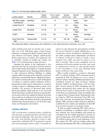
Table 1.6.  Commercially available lactate meters.

                                                                  Sample
                                           Range
  VetBooks.ir  Lactate analyzer  Mobility  (mmol/L)  Time to result   volume (μL)  Sample   Strip/cartridge/
                                                                                      sensor card
                                                                           type
                                                     (seconds)
              Stat Strip Lactate
                                                                             blood
               Connectivity a  Handheld    0.3–20       13         0.6     Whole      Strip
              Lactate Scout 4 a  Handheld  0.5–25       10         0.5     Whole      Strip
                                                                             blood
              Lactate Plus a  Handheld     0.3–25       13         0.7     Whole      Strip
                                                                             blood
              i-STAT b       Handheld      0.3–20      120         95      Whole      Cartridge
                                                                             blood
              Nova Prime Plus   Transportable  0.4–20   60       130–135   Whole      Sensor card
               Vet a                                                         blood
             a Nova Biomedical, Waltham, Massachusetts, USA;  Abbott Point of Care, Abbott Laboratories, Abbott Park, Illinois, USA.
                                             b
             often multifactorial and can involve one or more   return of color. Reasons for prolongation include,
             than one of the following: upper airway disease,   but are not limited to, shock, dehydration, vaso-
             pulmonary parenchymal disease, pleural space dis-  constriction, severe vasodilation, hypothermia, or
             ease, thoracic wall injury, primary cardiovascular   any other condition that compromises circulation
             disease, pain, stress, or anemia. The respiratory rate   or delivery of blood to tissues or leads to poor
             is normally counted as breaths per minute (see   vascular tone. CRTs can also be rapid (i.e. less
             Table 1.2 for normal rates in dogs and cats).  than 1 second). This is most commonly seen in
               Assessing the quality of the respirations is also   situations with supra-normal vascular tone such
             important and has the potential to provide informa-  as compensatory shock (i.e. the smooth muscle
             tion to help localize the problem to a specific location   in  the  vasculature  is very taut allowing rapid
             along the respiratory tract. Hearing crackles, wheezes,   return of blood to the tissue bed).
             or rales (abnormal clicking, bubbling, or rattling   What actually constitutes a normal or abnormal
             sounds) within the lungs can be indicative of primary   CRT is a subject of debate. CRT in humans was first
             pulmonary parenchymal disease. Significant inspira-  promoted in 1947 by Beecher as a way to assess a
             tory stridor (sound produced as a result of disruption   patient in shock. Initially, published parameters were
             of airflow) in a patient could be indicative of laryn-  considered  ‘normal’ (no signs of shock),  ‘definite
             geal paralysis, upper airway obstruction as a result of   slowing’ (slight to moderate shock), and ‘very slug-
             a polyp or mass, or collapsing trachea to name a few   gish’ (‘severe’ shock). In 1980, Champion and col-
             examples.  The presence of decreased lung sounds   leagues  incorporated  these  ranges  into  the  human
             during auscultation might lead one to be concerned   ‘Trauma Score’ and in so doing set the normal CRT
             about the presence of pleural effusion or pneumotho-  as less than 2 seconds despite a lack of scientific data
             rax. In cases with severe respiratory distress, decreased   to support this as being normal. Subsequently, stud-
             lung sounds are an indication for immediate thoraco-  ies related to CRT have determined that CRT actu-
             centesis to relieve the distress. If fluid is obtained and   ally varies with age and temperature (Schriger and
             analyzed from thoracocentesis, it can be both a diag-  Baraff, 1988) and that further research is needed to
             nostic and therapeutic intervention.        establish the true validity and usefulness of the CRT
                                                         as an accurate measure of circulatory status.
                                                           To date no studies evaluating CRT and its use as a
             Capillary refill time
                                                         predictor of perfusion or circulatory status in veteri-
             CRT is tested when a mucosal surface (usually the   nary medicine have been identified, and normal has
             oral mucosa) is pressed until the color leaves and   continued to be the initially proposed <2 seconds. As
             the tissue bed blanches. The time that it takes for   such, the importance of the CRT is somewhat unclear.
             the color (and therefore blood) to return to that   The authors propose that it is most important to seri-
             tissue bed is called the CRT. It is generally consid-  ally examine the CRT and note changes rather than
             ered to be prolonged when it takes >2 seconds for   the actual documented CRT. Also, evaluating the CRT


             Physical Examination and Point-of-care Testing                                   13