
Page 22 - Basic Monitoring in Canine and Feline Emergency Patients
P. 22
in the context of the remainder of the PE is more hepatic, or post-hepatic (biliary tree) abnormalities.
important than evaluating it on its own. Hyperemic/injected mucous membranes (also referred
VetBooks.ir to as ‘brick red’) can occur in some patients as a result
of septic conditions or endotoxemia, which cause
vasodilation. The bright red color is caused by the
Mucous membrane color
presence of blood pooling in the dilated capillaries in
Mucous membrane color is normally pink. Changes the gingiva. Mucous membranes can also have a blu-
in MM color can be influenced by changes in tissue ish color (i.e. cyanotic); this occurs as a result of the
perfusion, vasomotor tone (venoconstriction or presence of excessive deoxygenated blood (see
dilation), or the presence of deoxygenated hemo- Chapter 4 for more information).
globin or other pigments such as bilirubin (see Fig.
1.10). While assessing the color, it is important to
be aware that the ability to assess the MM color is Point-of-care blood testing
affected by gingival pigmentation, ambient lighting, Packed cell volume/total protein
and visual acuity, making assessment of MM color
variable between operators. Table 1.7 summarizes the information collected
Pale mucous membranes generally indicate vaso- from evaluating the PCV/TP.
constriction or anemia, both of which can have a A decreased PCV could indicate anemia as a
variety of causes. Assessment for causes of shock result of loss, destruction or lack of production of
based on other physical exam findings and measure- RBCs. An increased PCV could result from poly-
ment of PCV/TP should be prompted when pale MM cythemia or dehydration.
are found. Determination of body temperature and The presence of an increased TP is not pathogno-
glucose levels are also indicated as pallor also occurs monic for any particular disease process but could
in hypoglycemia and hypothermia. Yellow (icteric, be indicative of such disorders as dehydration,
jaundiced) MM may indicate pre-hepatic (hemolysis), inflammation, or even neoplasia. A decreased TP
(A) (B) (C)
(D) (E)
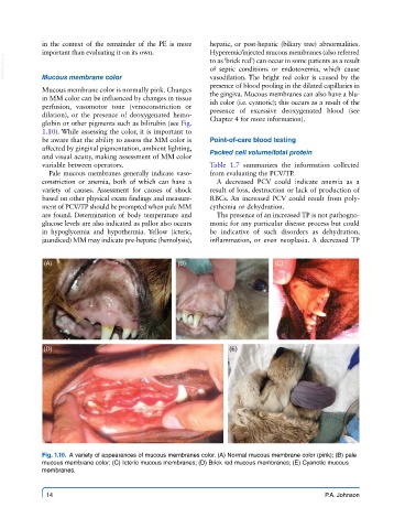
Fig. 1.10. A variety of appearances of mucous membranes color. (A) Normal mucous membrane color (pink); (B) pale
mucous membrane color; (C) Icteric mucous membranes; (D) Brick red mucous membranes; (E) Cyanotic mucous
membranes.
14 P.A. Johnson