
Page 23 - Basic Monitoring in Canine and Feline Emergency Patients
P. 23
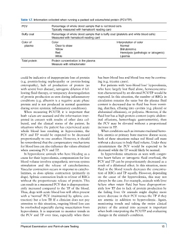
Table 1.7.  Information collected when running a packed cell volume/total protein (PCV/TP).

  VetBooks.ir  PCV             Percentage of whole blood sample that is red blood cells
                               Typically measured with hematocrit reading card
              Buffy coat
                               Percentage of whole blood sample that is buffy coat (platelets and white blood cells)
                               Measured with hematocrit reading card
              Color of         Color                                Interpretation of color
               plasma            Clear to straw                       Normal
                                 Yellow                               Bilirubinemia
                                 Red                                  Hemolysis (pathologic or iatrogenic)
                                 White                                Lipemia
              Total protein    Protein concentration in the plasma
                               Measure with refractometer


             could be indicative of inappropriate loss of protein   has been blood loss and blood loss may be continu-
             (e.g. protein-losing nephropathy or protein-losing   ing (e.g. trauma cases).
             enteropathy), lack of production of protein (as   For patients with ‘non-blood loss’ hypovolemia,
             with severe liver disease), iatrogenic dilution if fol-  who have largely lost fluid alone, hemoconcentra-
             lowing fluid therapy, or temporary downregulation   tion characterized by an elevated PCV/TP would be
             of protein production as seen during inflammatory   expected. In this situation, the number of RBCs in
             conditions (e.g. albumin is a negative acute phase   circulation remains the same but the plasma fluid
             protein and is not produced in normal quantities   content is decreased due to fluid loss from vomit-
             during severe systemic inflammatory conditions).  ing, diarrhea, effusing into cavities (e.g. pleural or
               When  measuring  PCV/TP,  it  is  important  that   abdominal effusions), or polyuria. However, if the
             both values are assessed and the information inter-  fluid lost has a high protein content (septic abdom-
             preted in concert with results of other data col-  inal effusions, hemorrhagic gastroenteritis), then
             lected, and the clinical status of the patient. In   the PCV may be elevated without a proportional
             situations where the patient has experienced acute   increase in TP.
             whole blood loss resulting in hypovolemia, the   When conditions such as immune-mediated hemo-
             PCV and TP would be expected to be decreased   lytic anemia or primary bone marrow disease occur,
             proportionally to one another. However, it should   both of these situations reduce red blood cell mass
             be remembered that the compensatory mechanisms   without a decrease in body fluid volume. Under these
             for blood loss can also influence the values obtained   circumstances the PCV would be expected to be
             when assessing PCV and TP.                  decreased while the TP would likely be normal.
               In hypovolemic animals who have bleeding as a   In hypervolemic situations as seen with conges-
             cause for their hypovolemia, compensation for low   tive heart failure or iatrogenic fluid overload, the
             blood volume involves sympathetic nervous system   PCV and TP can be proportionately decreased as a
             stimulation and the release of catecholamines.   result of a dilutional effect. In this situation, more
             Vascular contraction occurs in response to catecho-  fluid in the blood vessels decreases the concentra-
             lamines, as does splenic contraction (primarily in   tion of RBCs and TP equally. However, depending
             dogs). Splenic contraction leads to release of RBCs   on the cause of the hypervolemia, this may not
             without the proportionate release of protein. This   always be the case. For example, animals with liver
             can result in a measured PCV that is disproportion-  failure often retain fluid but have disproportion-
             ately increased compared to the TP of the blood.   ately low TP due to lack of protein production by
             Thus, dogs with acute blood loss will often present   the failing liver. Or animals might display more
             with a ‘normal’ PCV (maintained by splenic con-  severe decrease in their PCV versus the TP if they
             traction) but a low TP. If a clinician does not pay   are anemic in addition to hypervolemic.  Again,
             attention to this situation, ongoing blood loss can   monitoring trends and taking the entire clinical
             be overlooked especially during resuscitation from   picture of the animal into account is important
             hypovolemia. It is important to monitor trends in   when both interpreting the PCV/TP and evaluating
             the PCV and TP over time, especially when there   changes in the animal’s condition.


             Physical Examination and Point-of-care Testing                                   15