
Page 295 - Veterinary Toxicology, Basic and Clinical Principles, 3rd Edition
P. 295
262 SECTION | II Organ Toxicity
VetBooks.ir accumulation within glomerular capillary walls, mesan- (Hosseininejad and Hosseini, 2008). Lesions found in ani-
mals with CRF include kidneys that are small and irregular
gial dysfunction associated with matrix accumulation
in shape, with uneven capsular surfaces. On cut section,
and microaneurysm formation, and thrombosis due to
endothelial injury (Polzin, 2010). As the glomerulus pale streaks (fibrosis) may be seen within the interstitium
expands, podocytes are unable to maintain the integrity and the parenchyma may be gritty upon cutting due to min-
of slit diaphragms and focal denudation of GBM occurs, eralization and/or crystal deposition.
allowing leakage of larger proteins into the glomerular
filtrate (proteinuria). In addition to hemodynamically
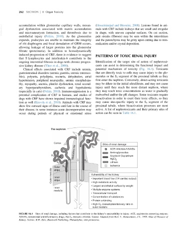
induced progression of CRF, there is evidence to suggest PATTERNS OF TOXIC RENAL INJURY
that T-lymphocytes and interleukin-6 contribute to the
ongoing interstitial fibrosis in dogs with chronic progres- Identification of the target site of action of nephrotoxi-
sive kidney disease (Yhee et al., 2008). cants can assist in determining the functional impact and
Clinical effects associated with CRF include uremia, potential mechanism of toxicity (Fig. 16.1). Toxicants
gastrointestinal disorders (uremic gastritis, uremic enteroco- that are directly toxic to cells may cause injury to the glo-
litis), polyuria, polydipsia, nocturia, dehydration, atrial merulus or the S 1 segment of the proximal tubule as they
hypertension, peripheral neuropathy, uremic encephalopa- first enter the nephron. Conversely, direct-acting toxicants
thy, myopathy, anemia, platelet dysfunction, renal second- may be dilute in the initial ultrafiltrate, and may not cause
ary hyperparathyroidism, cachexia and hypokalemia injury until they reach the more distant nephron, where
(especially in cats) (Polzin, 2010). Immunosuppression is a they may reach toxic concentrations as water is gradually
potential complication of CRF in humans, and studies of reabsorbed and/or the pH changes. Some toxicants require
dogs with CRF have shown impaired immunological func- bioactivation in order to exert their toxic effects, so they
tion as well (Kravola et al., 2010). Animals with CRF may may cause site-specific injury to the S 3 segment of the
show few outward signs of illness until late in the course of proximal tubule, where bioactivation processes are most
their disease; in some instances acute decompensation may active. A list of nephrotoxicants and their primary sites of
occur during periods of physical or emotional stress action can be seen in Table 16.1.
The nephron
S 1
Cortex S 2 Sites of renal damage
Medullary ray ACE inhibitors NSAIDs
Aminoglycosides
S 1 Acyclovir Cisplatinum
S HgCI 2
S 2 3 Lithium
Ischemia
Outer S 3
Outer medulla Inner Vulnerability of the kidney
stripe
• Important blood flow (1/4 cardiac output)
stripe
• High metabolic activity
• Largest endothelial surface by weight
Inner
medulla • Multiple enzyme systems
• Transcellular transport
• Concentration of substances
• Protein unbinding
• High O 2 consumption/delivery ratio in
outer medulla
FIGURE 16.1 Sites of renal damage, including factors that contribute to the kidney’s susceptibility to injury. ACE, angiotensin converting enzyme;
NSAIDs, nonsteroidal antiinflammatory drugs; HgCl 2 , mercuric chloride. Source: Adapted from Berl, T., Bonaventure, J.V., 1998. Atlas of Diseases of
Kidney. Schrier, R.W. (Ed.), Blackwell Publishing, Philadelphia, with permission.