
Page 514 - Veterinary Toxicology, Basic and Clinical Principles, 3rd Edition
P. 514
Salt Toxicity Chapter | 34 481
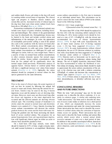
VetBooks.ir and sudden death. Excess salt intake in the dog will result serum sodium concentration is the first step in treatment
on an individual animal basis. This information can be
in vomiting within several hours of ingestion. The clinical
signs can progress to diarrhea, muscle tremors and
used to correct the free water deficit (FWD) in the animal,
seizure-like activity. Increased severity of clinical signs in based on the following formula:
the dog have been seen when serum sodium levels have
FWD ð1Þ 5 0:6 3 body weight ðkgÞ
been above 180 mEq/l (Barr et al., 2004).
3 ½ðmeasured serum Na=normal serum NaÞ 2 1
Postmortem examination of salt-poisoned animals may
include some degree of gastric irritation, including ulcera- Not more than 50% of the FWD should be replaced in
tion and hemorrhages. The content of the gastrointestinal the first 24 h with the remaining deficit replaced in the
tract may be abnormally dry. Histopathologic lesions may following 24 48 h. Serum sodium levels should be low-
be limited to the brain and include cerebral edema and ered at a rate of 0.5 1.0 mEq/L/h, with the slower rate
inflammation of the meninges. In swine, the appearance recommended for cases of chronic hypernatremia
of eosinophilic perivascular cuffing is seen if the animal (Schaer, 2000). In dogs with acute hypernatremia, the
dies early in the syndrome with the lesion not found after use of warm water enemas (6.6 11 mL/kg) repeated
48 h. Brain sodium concentrations above 2000 ppm are every 1 2 h has been suggested (Donaldson, 2003;
considered diagnostic in cattle and swine. Upper normal Howard, 2007). In acute hypernatremia without clinical
brain sodium concentrations are 1600 ppm for cattle and dehydration, the use of 5% dextrose solution in combina-
1800 ppm for swine, both on a wet weight basis. There is tion with a loop diuretic has been suggested at 3.7 mL/kg/h
a paucity of data on normal brain sodium concentrations to decrease serum sodium at 1 mEq/L/h (Barr et al.,
in other common domestic species but normal ranges 2004). Diuretics such as furosemide can be used to pre-
should be similar. Serum sodium concentrations taken vent the development of pulmonary edema during fluid
from the live animal will be significantly above the therapy. The use of slightly hypertonic intravenous fluids
normal ranges listed previously. Postmortem analysis of has been recommended to reduce the likelihood of cere-
aqueous humor, vitreous humor or cerebral spinal fluid bral edema developing. Intravenous fluids should be made
will show a significant increase over values from normal to approximate the serum sodium concentration of the ani-
animals (Osweiler et al., 1995). Optimally, the values mal, or the clinician may start with a solution containing
obtained should be compared to normal values for that 170 mEq/L of sodium and decrease this concentration as
species generated by the same laboratory. clinical signs improve (Angelos and Van Metre, 1999;
Niles, 2004). If brain edema is suspected, the use of man-
nitol, dexamethasone, or dimethyl sulfoxide may aid in
TREATMENT control.
Prior to the onset of clinical signs, the acute ingestion of
salt can best be treated by allowing the animal full CONCLUDING REMARKS
access to water and closely observing the animal for sev-
AND FUTURE DIRECTIONS
eral hours. Emetics may be used in the dog if known
ingestions occur and the animal is not yet showing clini- While the term “salt poisoning” may not be the most
cal signs. However, most cases are discovered long after accurate way to describe the above syndrome, it is cer-
the excess salt ingestion or the water deprivation has tainly in common usage. A similar and confusing term,
occurred and the affected animals are showing obvious “water intoxication,” has been used to describe the situa-
clinical signs. The overriding concept of treatment is to tion of excess water intake or infusion over a short period
slowly return the animal to normal water and electrolyte of time, which can dramatically decrease the serum
balance over a period of 2 3 days. Quickly lowering the sodium concentration and make the serum hypoosmolar.
serum sodium concentration will increase the osmotic Water intoxication has been used to describe the exacer-
gradient between the serum and the brain with water fol- bation of cerebral edema when the correction of hyperna-
lowing the gradient into the brain increasing the likeli- tremia occurs too quickly. Water intoxication has also
hood of severe cerebral edema. The prognosis for an been used to describe the brain swelling and seizure-like
animal hypernatremic from salt ingestion/water depriva- activity which occurs when a normal animal drinks
tion with significant clinical signs on either an acute or excessive amounts of water over a short period of time.
chronic basis should be guarded at best. In addition to the possible neurological effects, hemolysis
On a herd basis with large animals, water intake has also been described (Middleton et al., 1997). In the
should be limited to 0.5% of body weight at hourly inter- above situations, acute and dramatic osmotic changes are
vals until normal hydration is accomplished. Monitoring the cause.