
Page 688 - Veterinary Toxicology, Basic and Clinical Principles, 3rd Edition
P. 688
Alcohols and Glycols Chapter | 49 653
VetBooks.ir also increase serum osmolality. Serum osmolality is
increased within an hour of EG ingestion, increasing
in parallel with serum EG concentrations (Dial et al.,
1994a,b). When measured serum osmolality (by osmome-
try) is compared to calculated serum osmolality, the differ-
ence is referred to as the osmole or osmolal gap. Normal
serum osmolality is 280 310 mOsm/kg, and the normal
osmole gap is less than 10 mOsm/kg. Serum osmolality as
high as 450 mOsm/kg and an osmole gap as high as
150 mOsm/kg may be seen 3 h after ingestion, depending
on the quantity of antifreeze ingested (Jacobsen et al.,
1982b; Grauer et al., 1984). Both the gap and the measured
osmolality may remain significantly high for approximately
18 h after ingestion. Multiplication of the osmole gap
by five yields an approximate serum EG concentration in FIGURE 49.3 Calcium oxalate monohydrate crystals (polarized light)
mg/dL (Burkhart and Kulig, 1990). Each 100 mg/dL incre- from a dog with EG toxicosis.
ment increase in EG concentration contributes approxi-
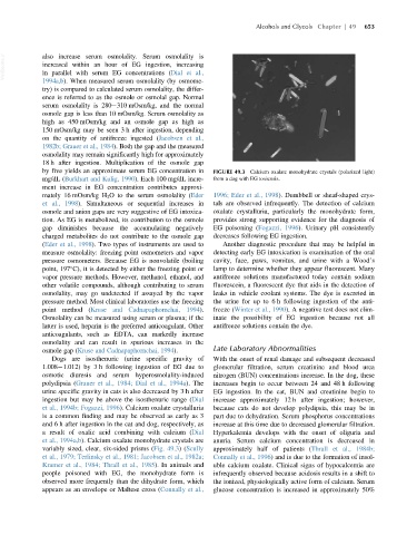
mately 16 mOsm/kg H 2 O to the serum osmolality (Eder 1996; Eder et al., 1998). Dumbbell or sheaf-shaped crys-
et al., 1998). Simultaneous or sequential increases in tals are observed infrequently. The detection of calcium
osmole and anion gaps are very suggestive of EG intoxica- oxalate crystalluria, particularly the monohydrate form,
tion. As EG is metabolized, its contribution to the osmole provides strong supporting evidence for the diagnosis of
gap diminishes because the accumulating negatively EG poisoning (Fogazzi, 1996). Urinary pH consistently
charged metabolites do not contribute to the osmole gap decreases following EG ingestion.
(Eder et al., 1998). Two types of instruments are used to Another diagnostic procedure that may be helpful in
measure osmolality: freezing point osmometers and vapor detecting early EG intoxication is examination of the oral
pressure osmometers. Because EG is nonvolatile (boiling cavity, face, paws, vomitus, and urine with a Wood’s
point, 197 C), it is detected by either the freezing point or lamp to determine whether they appear fluorescent. Many
vapor pressure methods. However, methanol, ethanol, and antifreeze solutions manufactured today contain sodium
other volatile compounds, although contributing to serum fluorescein, a fluorescent dye that aids in the detection of
osmolality, may go undetected if assayed by the vapor leaks in vehicle coolant systems. The dye is excreted in
pressure method. Most clinical laboratories use the freezing the urine for up to 6 h following ingestion of the anti-
point method (Kruse and Cadnapaphornchai, 1994). freeze (Winter et al., 1990). A negative test does not elim-
Osmolality can be measured using serum or plasma; if the inate the possibility of EG ingestion because not all
latter is used, heparin is the preferred anticoagulant. Other antifreeze solutions contain the dye.
anticoagulants, such as EDTA, can markedly increase
osmolality and can result in spurious increases in the
osmole gap (Kruse and Cadnapaphornchai, 1994). Late Laboratory Abnormalities
Dogs are isosthenuric (urine specific gravity of With the onset of renal damage and subsequent decreased
1.008 1.012) by 3 h following ingestion of EG due to glomerular filtration, serum creatinine and blood urea
osmotic diuresis and serum hyperosmolality-induced nitrogen (BUN) concentrations increase. In the dog, these
polydipsia (Grauer et al., 1984; Dial et al., 1994a). The increases begin to occur between 24 and 48 h following
urine specific gravity in cats is also decreased by 3 h after EG ingestion. In the cat, BUN and creatinine begin to
ingestion but may be above the isosthenuric range (Dial increase approximately 12 h after ingestion; however,
et al., 1994b; Fogazzi, 1996). Calcium oxalate crystalluria because cats do not develop polydipsia, this may be in
is a common finding and may be observed as early as 3 part due to dehydration. Serum phosphorus concentrations
and 6 h after ingestion in the cat and dog, respectively, as increase at this time due to decreased glomerular filtration.
a result of oxalic acid combining with calcium (Dial Hyperkalemia develops with the onset of oliguria and
et al., 1994a,b). Calcium oxalate monohydrate crystals are anuria. Serum calcium concentration is decreased in
variably sized, clear, six-sided prisms (Fig. 49.3)(Scully approximately half of patients (Thrall et al., 1984b;
et al., 1979; Terlinsky et al., 1981; Jacobsen et al., 1982a; Connally et al., 1996) and is due to the formation of insol-
Kramer et al., 1984; Thrall et al., 1985). In animals and uble calcium oxalate. Clinical signs of hypocalcemia are
people poisoned with EG, the monohydrate form is infrequently observed because acidosis results in a shift to
observed more frequently than the dihydrate form, which the ionized, physiologically active form of calcium. Serum
appears as an envelope or Maltese cross (Connally et al., glucose concentration is increased in approximately 50%