
Page 1229 - Small Animal Internal Medicine, 6th Edition
P. 1229
CHAPTER 69 Disorders of the Joints 1201
Clinical abnormalities develop 3 months to 7 years after affected joints has been unrewarding, although the virus can
infection and typically consist of vague signs, including be found in the oropharynx of some infected cats. The poly-
VetBooks.ir weight loss, lymphadenopathy, and splenomegaly. Hyper- arthritis in these kittens is likely to be immune complex
mediated (reactive polyarthritis) rather than infectious.
globulinemia, hypoalbuminemia, and proteinuria are
expected. Polyarthritis causing lameness and exercise intol-
erance is common. Many affected dogs will have erosive
disease with radiographic evidence of periarticular lysis and NONINFECTIOUS POLYARTHRITIS:
periosteal proliferation. Diagnosis is made when organisms NONEROSIVE
are identified within macrophages in lymph node or splenic
aspirates or in joint fluid (see Chapter 98). Noninfectious inflammatory (immune-mediated) joint dis-
eases are very common in the dog but rare in the cat. IMPA
FUNGAL ARTHRITIS is classified as either erosive or nonerosive on the basis of
Fungal infection of the joints is very rare. When it does the presence or absence of radiographically evident joint
occur, it is usually as an extension of fungal osteomyelitis destruction. Erosive disorders are very rare (<1% of canine
caused by Coccidioides immitis, Blastomyces dermatitidis, or polyarthritis cases). Nonerosive IMPA is believed to be
Cryptococcus neoformans. More commonly a reactive, immu- mediated through immune complex formation and deposi-
nologically mediated, culture-negative polyarthritis occurs tion in the synovial membrane. Immune-mediated nonero-
in dogs and cats with systemic fungal infections. sive polyarthritis occurs secondary to antigenic stimulation
from chronic infection, neoplasia, or drugs (i.e., reactive
polyarthritis), as an idiopathic syndrome, and as a feature
VIRAL ARTHRITIS of systemic lupus erythematosus (SLE). Breed-associated
Calicivirus syndromes of polyarthritis or polyarthritis/meningitis or
Natural calicivirus infection and attenuated live calicivirus polyarthritis/myositis also exist and are thought to have a
vaccination have been associated with development of tran- genetic basis.
sient polyarthritis in 6- to 12-week-old kittens. Clinical signs
include lameness, stiffness, and fever that usually resolve REACTIVE POLYARTHRITIS
spontaneously after 2 to 4 days (Fig. 69.4). Some kittens go Reactive polyarthritis accounts for some 25% of all nonero-
on to develop overt calicivirus infection, with glossal and sive IMPAs cases and has been documented in association
palatine vesicles or ulcers and signs of upper respiratory tract with chronic bacterial, fungal, or rickettsial infections; neo-
disease. Synovial fluid analysis reveals a mildly to greatly plasia; and drug administration. When it occurs secondary
increased nucleated cell count, with small mononuclear cells to chronic infection, the joints themselves are not infected,
and macrophages predominating, some of which contain but immune complex deposition causes immune mediated
phagocytosed neutrophils. Two specific strains of calicivirus synovitis. Reactive polyarthritis has been documented in
have been implicated. Attempts to isolate the virus from dogs with endocarditis, foreign body abscesses or granulo-
mas, diskospondylitis, heartworm disease, pancreatitis, pros-
tatitis, pyelonephritis, pneumonia, other chronic bacterial
and rickettsial infections, and a variety of tumors (Fig. 69.5).
Drugs that have been implicated in causing reactive polyar-
thritis include sulfadiazine-trimethoprim, phenobarbital,
erythropoietin, penicillin, cephalexin, and routine vaccina-
tions. Rarely, gastrointestinal disorders such as inflammatory
bowel disease, salmonellosis, and chronic active hepatitis
have also been associated with reactive polyarthritis.
Because many animals with reactive polyarthritis have
vague or minimal clinical signs referable to their underlying
disease, they may be presented for veterinary evaluation
when their joint inflammation makes them reluctant to walk.
Therefore it is important to perform a thorough physical
examination of every animal with polyarthritis and obtain a
complete history regarding medication administration and
the presence or absence of systemic signs. Once infectious
causes of polyarthritis have been eliminated, screening tests
(i.e., complete blood count [CBC], biochemical panel, uri-
nalysis, thoracic and abdominal radiography, abdominal
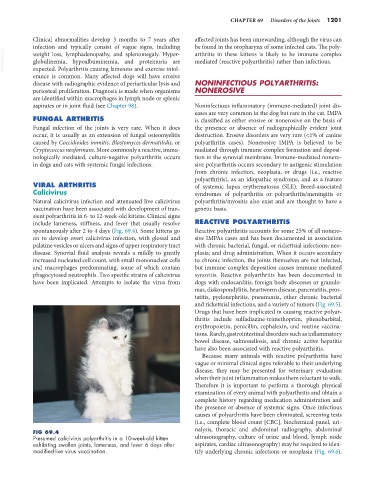
FIG 69.4
Presumed calicivirus polyarthritis in a 10-week-old kitten ultrasonography, culture of urine and blood, lymph node
exhibiting swollen joints, lameness, and fever 6 days after aspirates, cardiac ultrasonography) may be required to iden-
modified-live virus vaccination. tify underlying chronic infections or neoplasia (Fig. 69.6).