
Page 1234 - Small Animal Internal Medicine, 6th Edition
P. 1234
1206 PART X Joint Disorders
LYMPHOPLASMACYTIC SYNOVITIS time (months to years), with distal joints most severely
Lymphoplasmacytic synovitis is present in some dogs with affected.
VetBooks.ir partial and complete tears of the cranial cruciate ligament, Etiology
but the relationship between the immune-mediated response
and the ligament rupture is uncertain. Partial tears or rup-
understood. Antibodies directed against IgG (i.e., rheuma-
tures of the cruciate ligament commonly initiate an inflam- The pathogenesis of canine RA-like polyarthritis is poorly
matory reaction directed against the collagen of the ligament, toid factors [RFs]) form and complex with IgG within the
resulting in mildly inflammatory synovial fluid and synovial synovium. This results in complement activation and the
fluid antibodies directed against type 1 and type 2 collagen. chemotactic attraction of plasma cells, lymphocytes, and
An alternative theory is that lymphoplasmacytic synovitis is neutrophils into the joint fluid. The synovial membrane
a primary immune-mediated disorder that causes joint laxity thickens and develops a fibrous, vascular granulation tissue
and instability, eventually leading to rupture of the cranial (pannus) that invades articular cartilage, tendons, ligaments,
cruciate ligament. Some investigators have estimated that and subchondral bone. Proteolytic enzymes are released that
perhaps as many as 10% to 25% of cruciate ruptures in dogs erode the articular cartilage and subchondral bone, leading
are caused by this immunologic disorder, but this is a con- to joint collapse and radiographically visible “punched-out”
troversial claim. subchondral bone lesions. Articular and periarticular inflam-
Dogs diagnosed with lymphoplasmacytic synovitis are mation and instability lead to joint subluxation and luxation,
the same dogs typically presented for cruciate ligament resulting in joint deformity.
rupture, with Rottweilers, Newfoundlands, Staffordshire Bull
Terriers, and Labrador Retrievers most commonly affected. Clinical Features
Clinical signs are limited to acute or chronic lameness Affected dogs initially have signs indistinguishable from
involving one or both stifles. Cruciate ligament rupture at those of other forms of polyarthritis. A low-grade fever,
the time of diagnosis may be partial or complete, and often depression, anorexia, and reluctance to exercise are common.
there is no history of trauma. Arthroscopy or magnetic reso- Joint-related clinical signs such as joint pain and stiff gait are
nance imaging (MRI) may be required to confirm the diag- prominent. Signs may be sporadic initially, and stiffness is
nosis of partial rupture. Affected animals are in good body generally worse after rest and improves with mild exercise.
condition and are not systemically ill; CBC is normal. Syno- The joints may appear normal or be swollen and painful. The
vial fluid is thin and turbid, with an increased nucleated cell joints most commonly affected are the carpi, hocks, and
count (5000-20,000 cells/µL, but occasionally > 200,000/µL). phalanges, although stifles, elbows, and shoulders can also
Lymphocytes and plasma cells predominate (60%-90%) in be affected. As the disease progresses, clinical examination
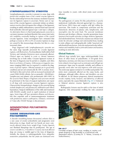
the synovial fluid. Biopsy of ligament and synovium should reveals crepitus, laxity, luxation, and deformity of affected
be performed at the time of surgical exploration and repair joints, especially the carpus bilaterally (Fig. 69.7). Unilateral
in all dogs with nontraumatic cruciate ligament ruptures. or bilateral anterior cruciate ligament rupture is also
Characteristic histopathologic changes in the synovial lining common.
include lymphocytic and plasmacytic infiltration and villous Radiographic features may be subtle at the time of initial
hyperplasia. Surgical stabilization of the stifle and treatment diagnosis, with intracapsular swelling the only consistent
with NSAIDs usually result in rapid resolution of clinical
signs. Some dogs will have persistent effusion and discom-
fort that responds well to immunosuppressive treatment
with prednisone and/or azathioprine, initiated a minimum
of 3 days after NSAID therapy is discontinued.
NONINFECTIOUS POLYARTHRITIS:
EROSIVE
CANINE RHEUMATOID-LIKE
POLYARTHRITIS
A disorder resembling human rheumatoid arthritis (RA) is
a rare cause of erosive polyarthritis and progressive joint
destruction in dogs. Small and toy breeds are most com-
monly affected (mean body weight 8 kg). Cocker Spaniels
and Shetland Sheepdogs may be predisposed. The age of FIG 69.7
onset is variable (i.e., 9 months to 13 years), but most affected Complete collapse of both carpi, resulting in luxation and
dogs are young or middle-aged at the time of diagnosis. severe distortion of the forelimbs in a Dachshund with
Initially, the disease is indistinguishable from idiopathic rheumatoid arthritis. (Courtesy Dr. D. Haines, University of
nonerosive polyarthritis, but the joints are destroyed over Saskatchewan.)