
Page 1235 - Small Animal Internal Medicine, 6th Edition
P. 1235
CHAPTER 69 Disorders of the Joints 1207
VetBooks.ir
A B
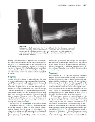
FIG 69.8
Radiographs of both carpal joints of a 9-year-old female Shih Tzu. Both carpi are severely
deformed secondary to erosive rheumatoid-like polyarthritis. The intercarpal spaces have
thinned laterally, and there are focal radiolucent cyst-like areas of subchondral bone
destruction and regional soft tissue swelling. There is dislocation of the radius and ulna
from the carpus bilaterally.
finding. Later, characteristic changes consist of focal, irregu- lymphocytes, plasma cells, macrophages, and neutrophils.
lar, radiolucent, cyst-like areas of subchondral bone destruc- Culture of the synovial biopsy is negative. RA is diagnosed
tion (Fig. 69.8); joint space collapse; and joint subluxation on the basis of the typical clinical findings and radiographic
and luxation. Erosive changes are common in the carpus at features, characteristic synovial fluid features, a positive RF
the time of diagnosis (75% of affected dogs) and are evident test result, and the typical histopathologic changes seen in a
radiographically in all affected dogs within 1 year of diagno- synovial biopsy specimen.
sis. Whenever RA is suspected, carpi should be radiographed
bilaterally. Treatment
Early treatment of RA is important to prevent irreversible
Diagnosis changes and progressive disease. Medical treatment usually
RA-like polyarthritis should be suspected in any dog with includes immunosuppressive drugs and chondroprotective
erosive polyarthritis once infectious causes have been elimi- agents. Initially, most dogs are treated with oral prednisone
nated. The synovial fluid in affected joints is thin, cloudy, and (2-4 mg/kg q24h for 14 days, then 1-2 mg/kg q24h for 14
hypercellular, with an increased TNCC similar to dogs with days) and azathioprine (2.2 mg/kg PO q24h), administered
nonerosive idiopathic IMPA (mean 7200 WBCs/µL). Neu- as described for the treatment of refractory idiopathic noner-
trophils are usually the predominant cell (20%-95%; average osive polyarthritis. Oral chondroprotective agents (see Table
74%), but mononuclear cells may sometimes predominate. 69.1) should be administered concurrently. Subjective
Lymphocyte numbers are often increased in synovial fluid improvement has also been observed in dogs receiving
from dogs with erosive polyarthritis. Culture of synovial injectable chondroprotective agents (e.g., Adequan).
fluid is negative. Whenever possible, the synovial fluid If there is a good response to treatment, based on both
should be collected during a period when the dog is most resolution of clinical signs and synovial fluid inflammation,
symptomatic, because the cyclical nature of the disease occa- the glucocorticoid dose should be decreased to 1 to 2 mg/kg
sionally makes diagnosis difficult. orally every 48 hours, and treatment with azathioprine is
Serologic tests for circulating RF are positive in 20% to continued. If the response to treatment is inadequate after 1
70% of affected dogs (see Chapter 68). Weak false-positive month of treatment with glucocorticoids and azathioprine,
results are common in dogs with other systemic inflam- more aggressive immunosuppressive therapy should be con-
matory diseases. Synovial biopsy may help establish the sidered (see Table 69.2). Few published data exist regarding
diagnosis, revealing synovial thickening, hyperplasia, and treatment of RA in dogs, so choice of immunosuppressive
proliferation with pannus formation. The pannus is com- agents is usually based on individual clinical experience and
posed primarily of proliferating activated synoviocytes, response to therapy. Leflunomide has been reported to be