
Page 1246 - Small Animal Internal Medicine, 6th Edition
P. 1246
1218 PART XI Immune-Mediated Disorders
dogs with IMPA had increased CRP concentrations at the immunofluorescent staining. In the case of immunoperoxi-
time of diagnosis; 82% of dogs demonstrated a significant dase peroxide, when a substrate is added in the presence of
VetBooks.ir decrease in CRP concentrations within 5 days of corticoste- hydrogen peroxide, deposition of a brown color can be visu-
alized with the light microscope. Tissue samples for immuno-
roid therapy. CRP concentrations measured at 1 to 2 weeks
following initiation of treatment were predictive that contin-
Routinely fixed tissue can be used for immunohistochemis-
ued treatment with immune-suppressive medications would fluorescence testing should be collected in Michel’s medium.
still be required to manage IMPA at the 6-month follow up. try. Common uses for immunofluorescence staining include
CRP concentrations also correlated with resolution of clini- evaluation of renal biopsies in dogs with suspected glomeru-
cal signs by 1 to 2 weeks following initiation of corticosteroid lonephritis, detection of antibodies directed against mega-
therapy. Therefore, although measurement of CRP is not karyocytic cells in the bone marrow, and evaluation of skin
useful for diagnosis of IMPA, it should be considered as a biopsies from patients with suspected immune-mediated
monitoring tool in lieu of repeated arthrocentesis in dogs skin disease.
that are clinically responding well to treatment.
AUTOIMMUNE PANELS
IMMUNOFLUORESCENCE AND Many laboratories offer an immune panel that typically
IMMUNOHISTOCHEMISTRY includes a CBC and platelet count, Coombs test, ANA, and
In many type II and type III immune-mediated diseases, RF. It would be unusual for all these tests to be appropriate
the presence of antibody in fixed tissues (e.g., kidney, skin) in an individual patient (Table 71.1). In addition to the cost
can be detected by immunofluorescence or immunoperoxi- of running such a panel, the significance of a positive test
dase techniques. Numerous variations on these methods may be difficult to determine in patients in which the test
exist, but, in general, sections of tissue are labeled with a was initially not indicated, and some of these tests can be
primary antibody (e.g., rabbit anti–dog IgG) and then a positive in dogs and cats with infectious diseases. For these
secondary antibody is added (e.g., anti–rabbit IgG), which reasons the clinician is encouraged to pick individual tests
has been conjugated to either fluorescein or the enzyme rather than automatically choosing an autoimmune panel in
peroxidase. If antibodies are present in the tissue sample, a dog or cat with suspected autoimmune or immune-
apple green fluorescence is seen under ultraviolet light with mediated disease.
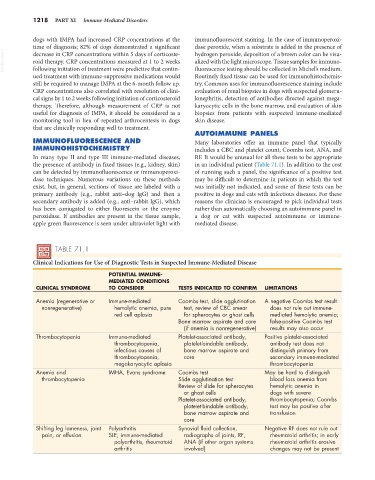
TABLE 71.1
Clinical Indications for Use of Diagnostic Tests in Suspected Immune-Mediated Disease
POTENTIAL IMMUNE-
MEDIATED CONDITIONS
CLINICAL SYNDROME TO CONSIDER TESTS INDICATED TO CONFIRM LIMITATIONS
Anemia (regenerative or Immune-mediated Coombs test, slide agglutination A negative Coombs test result
nonregenerative) hemolytic anemia, pure test, review of CBC smear does not rule out immune-
red cell aplasia for spherocytes or ghost cells mediated hemolytic anemia;
Bone marrow aspirate and core false-positive Coombs test
(if anemia is nonregenerative) results may also occur
Thrombocytopenia Immune-mediated Platelet-associated antibody, Positive platelet-associated
thrombocytopenia, platelet-bindable antibody, antibody test does not
infectious causes of bone marrow aspirate and distinguish primary from
thrombocytopenia, core secondary immune-mediated
megakaryocytic aplasia thrombocytopenia
Anemia and IMHA, Evans syndrome Coombs test May be hard to distinguish
thrombocytopenia Slide agglutination test blood loss anemia from
Review of slide for spherocytes hemolytic anemia in
or ghost cells dogs with severe
Platelet-associated antibody, thrombocytopenia; Coombs
platelet-bindable antibody, test may be positive after
bone marrow aspirate and transfusion
core
Shifting leg lameness, joint Polyarthritis Synovial fluid collection, Negative RF does not rule out
pain, or effusion SLE, immune-mediated radiographs of joints, RF, rheumatoid arthritis; in early
polyarthritis, rheumatoid ANA (if other organ systems rheumatoid arthritis erosive
arthritis involved) changes may not be present