
Page 1287 - Small Animal Internal Medicine, 6th Edition
P. 1287
CHAPTER 74 Cytology 1259
desmosomes), forming clusters or sheets. Individual cells are
easily identifiable and are round or polygonal; both nucleus
VetBooks.ir and cytoplasm are well differentiated (i.e., the nucleus is
small and has heavily clumped chromatin). Most cells in
Romanowsky-stained smears have blue cytoplasm and
round to oval nuclei.
Mesenchymal Tissues
Cells from mesenchymal tissues (e.g., fibroblasts, fibrocytes,
chondroblasts) are difficult to obtain in routine FNA mate-
rial or tissue scrapings because they are usually surrounded
by intercellular matrix. Mesenchymal cells are typically
spindle-shaped, polygonal, or oval and have irregular nuclei;
cytoplasmic boundaries are usually indistinct, and cell
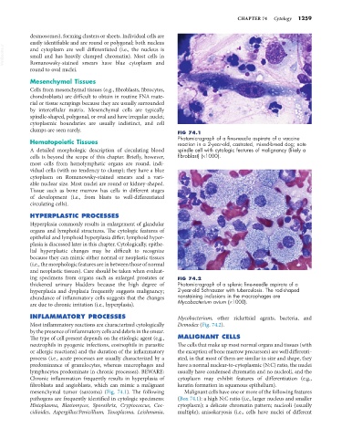
clumps are seen rarely. FIG 74.1
Photomicrograph of a fine-needle aspirate of a vaccine
Hematopoietic Tissues reaction in a 2-year-old, castrated, mixed-breed dog; note
A detailed morphologic description of circulating blood spindle cell with cytologic features of malignancy (likely a
cells is beyond the scope of this chapter. Briefly, however, fibroblast) (×1000).
most cells from hemolymphatic organs are round, indi-
vidual cells (with no tendency to clump); they have a blue
cytoplasm on Romanowsky-stained smears and a vari-
able nuclear size. Most nuclei are round or kidney-shaped.
Tissue such as bone marrow has cells in different stages
of development (i.e., from blasts to well-differentiated
circulating cells).
HYPERPLASTIC PROCESSES
Hyperplasia commonly results in enlargement of glandular
organs and lymphoid structures. The cytologic features of
epithelial and lymphoid hyperplasia differ; lymphoid hyper-
plasia is discussed later in this chapter. Cytologically, epithe-
lial hyperplastic changes may be difficult to recognize
because they can mimic either normal or neoplastic tissues
(i.e., the morphologic features are in between those of normal
and neoplastic tissues). Care should be taken when evaluat-
ing specimens from organs such as enlarged prostates or FIG 74.2
thickened urinary bladders because the high degree of Photomicrograph of a splenic fine-needle aspirate of a
hyperplasia and dysplasia frequently suggests malignancy; 2-year-old Schnauzer with tuberculosis. The rod-shaped
abundance of inflammatory cells suggests that the changes nonstaining inclusions in the macrophages are
are due to chronic irritation (i.e., hyperplasia). Mycobacterium avium (×1000).
INFLAMMATORY PROCESSES Mycobacterium, other rickettsial agents, bacteria, and
Most inflammatory reactions are characterized cytologically Demodex (Fig. 74.2).
by the presence of inflammatory cells and debris in the smear.
The type of cell present depends on the etiologic agent (e.g., MALIGNANT CELLS
neutrophils in pyogenic infections, eosinophils in parasitic The cells that make up most normal organs and tissues (with
or allergic reactions) and the duration of the inflammatory the exception of bone marrow precursors) are well differenti-
process (i.e., acute processes are usually characterized by a ated, in that most of them are similar in size and shape, they
predominance of granulocytes, whereas macrophages and have a normal nuclear-to-cytoplasmic (N:C) ratio, the nuclei
lymphocytes predominate in chronic processes). BEWARE: usually have condensed chromatin and no nucleoli, and the
Chronic inflammation frequently results in hyperplasia of cytoplasm may exhibit features of differentiation (e.g.,
fibroblasts and angioblasts, which can mimic a malignant keratin formation in squamous epithelium).
mesenchymal tumor (sarcoma) (Fig. 74.1). The following Malignant cells have one or more of the following features
pathogens are frequently identified in cytologic specimens: (Box 74.1): a high N:C ratio (i.e., larger nucleus and smaller
Histoplasma, Blastomyces, Sporothrix, Cryptococcus, Coc- cytoplasm); a delicate chromatin pattern; nucleoli (usually
cidioides, Aspergillus/Penicillium, Toxoplasma, Leishmania, multiple); anisokaryosis (i.e., cells have nuclei of different