
Page 1354 - Small Animal Internal Medicine, 6th Edition
P. 1354
1326 PART XII Oncology
considered relatively rare and appears to be more common Once a presumptive radiographic diagnosis has been
in Greyhounds, where approximately 15% of the patients established, and if the owners are contemplating treatment,
VetBooks.ir present due to a pathologic fracture. Physical examination thoracic radiographs or CT should be obtained to determine
the extent of the disease. The authors generally obtain three
usually reveals a painful swelling in the affected area, with
radiographic views of the thorax and do not perform skeletal
or without soft tissue involvement or pathologic fracture.
radiographic surveys (or radionuclide bone scan) unless
Diagnosis there are clinical signs that warrant this investigation (i.e.,
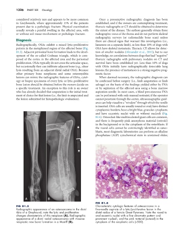
Radiographically, OSAs exhibit a mixed lytic-proliferative lameness on a separate limb), as less than 10% of dogs with
pattern in the metaphyseal region of the affected bone (Fig. OSA have skeletal metastasis. Thoracic CT allows for detec-
81.5). Adjacent periosteal bone formation leads to the devel- tion of smaller nodules (Alexander et al., 2012), but to our
opment of the so-called Codman triangle, which is com- knowledge, no correlations between dogs that had “negative”
posed of the cortex in the affected area and the periosteal thoracic radiographs with pulmonary nodules on CT and
proliferation. OSAs typically do not cross the articular space, survival have been established yet. Less than 10% of dogs
but occasionally they can infiltrate adjacent bone (e.g., ulnar with OSAs initially have radiographically detectable lung
lysis resulting from an adjacent distal radial OSA). Because lesions; the presence of metastases is a strong negative prog-
other primary bone neoplasms and some osteomyelitis nostic factor.
lesions can mimic the radiographic features of OSAs, cytol- When deemed necessary, the radiographic diagnosis can
ogy or biopsy specimens of every lytic or lytic-proliferative be confirmed before surgery (i.e., limb amputation or limb
bone lesion should be obtained before the owners decide on salvage) on the basis of the findings yielded either by FNA
a specific treatment. An exception to this rule is an owner or by aspiration of the affected area using a bone marrow
who has already decided that amputation is the initial treat- aspiration needle. In most cases, a blind percutaneous FNA
ment of choice for that lesion (i.e., the limb is amputated and can be performed with only manual restraint; if the operator
the lesion submitted for histopathologic evaluation). cannot penetrate through the cortex, ultrasonographic guid-
ance can help visualize a “window” through which the needle
is inserted. OSA cells are usually round or oval; have distinct
cytoplasmic borders; have a bright blue, granular cytoplasm;
and have eccentric nuclei with or without nucleoli (Fig.
81.6). Osteoclast-like multinucleated giant cells are common,
and there is frequently pink amorphous material (osteoid)
in the background or in the cytoplasm of the osteoblasts. If
the round cells cannot be convincingly identified as osteo-
blasts, most diagnostic laboratories can perform an alkaline
phosphatase (ALP) cytochemical stain in unstained slides;
A B
FIG 81.6
FIG 81.5 Characteristic cytologic features of osteosarcoma in a
Radiographic appearance of an osteosarcoma in the distal fine-needle aspirate of a lytic/proliferative lesion in the
tibia of a Greyhound; note the lytic and proliferative distal radius of a female Great Pyrenees. Note the round to
changes characteristic of this neoplasm (A). Radiographic oval eccentric nuclei with a fine chromatin pattern and
appearance of a distal radial osteosarcoma with massive prominent nucleoli, and the pink material (osteoid) in the
neoplastic new bone formation in a Mastiff (B). cytoplasm of the neoplastic cells (×500).