
Page 1359 - Small Animal Internal Medicine, 6th Edition
P. 1359
CHAPTER 81 Selected Neoplasms in Dogs and Cats 1331
Buffy coat smears to search for circulating mast cells As a general rule, in dogs with solitary MCTs in a location
are not clinically useful. Interestingly, circulating mast cells amenable to wide surgical excision and no negative prognos-
VetBooks.ir are more common in dogs with diseases other than MCTs; tic indicators, routine staging diagnostics (with the excep-
tion of regional lymph node aspiration) are not considered
most dogs with mastocythemia have inflammatory disor-
ders, regenerative anemia, tumors other than MCTs, or
indication of “aggressive” histopathology (i.e., high grade)
trauma. Circulating mast cells can frequently be recognized mandatory and can be performed postoperatively if there is
in the graphics of flow cytometry-based hematology ana- (Table 81.1).
lyzers (Fig. 81.8). Cytologic evaluation of a bone marrow
aspirate may therefore be more beneficial for staging pur- Treatment and Prognosis
poses, when deemed appropriate. On the basis of all these As discussed previously, it is imperative to know whether the
facts, the appropriate staging procedures in dogs with MCTs mass the clinician is preparing to excise is a MCT before
remain controversial. The authors do not use buffy coat surgery, because this information is useful when discussing
smears or bone marrow aspirates routinely in dogs with treatment options with the family and when planning the
MCT and a normal CBC; if cytopenias or leukoerythro- treatment strategy. Dogs with MCT can be treated with
blastic reactions are present, a bone marrow aspirate may surgery, radiotherapy, chemotherapy, molecular-targeted
be performed. therapy, or a combination of these. However, the first two
A B
C
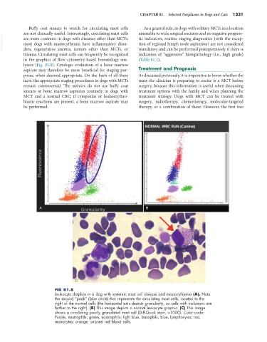
FIG 81.8
Leukocyte dotplots in a dog with systemic mast cell disease and mastocythemia (A). Note
the second “peak” (blue circle) that represents the circulating mast cells, located to the
right of the normal cells (the horizontal axis depicts granularity, so cells with inclusions are
further to the right). (B) This image depicts a normal leukocyte graphic. (C) This image
shows a circulating poorly granulated mast cell (Diff-Quick stain, ×1000). Color code:
Purple, neutrophils; green, eosinophils; light blue, basophils; blue, lymphocytes; red,
monocytes; orange, unlysed red blood cells.