
Page 652 - Small Animal Internal Medicine, 6th Edition
P. 652
624 PART IV Hepatobiliary and Exocrine Pancreatic Disorders
VetBooks.ir
A B
C D
E
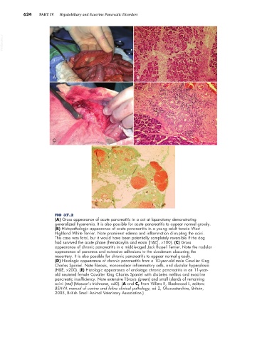
FIG 37.2
(A) Gross appearance of acute pancreatitis in a cat at laparotomy demonstrating
generalized hyperemia. It is also possible for acute pancreatitis to appear normal grossly.
(B) Histopathologic appearance of acute pancreatitis in a young adult female West
Highland White Terrier. Note prominent edema and inflammation disrupting the acini.
This case was fatal, but it would have been potentially completely reversible if the dog
had survived the acute phase (hematoxylin and eosin [H&E], ×100). (C) Gross
appearance of chronic pancreatitis in a middle-aged Jack Russell Terrier. Note the nodular
appearance of pancreas and extensive adhesions to the duodenum obscuring the
mesentery. It is also possible for chronic pancreatitis to appear normal grossly.
(D) Histologic appearance of chronic pancreatitis from a 10-year-old male Cavalier King
Charles Spaniel. Note fibrosis, mononuclear inflammatory cells, and ductular hyperplasia
(H&E, ×200). (E) Histologic appearance of end-stage chronic pancreatitis in an 11-year-
old neutered female Cavalier King Charles Spaniel with diabetes mellitus and exocrine
pancreatic insufficiency. Note extensive fibrosis (green) and small islands of remaining
acini (red) (Masson’s trichrome, ×40). (A and C, From Villiers E, Blackwood L, editors:
BSAVA manual of canine and feline clinical pathology, ed 2, Gloucestershire, Britain,
2005, British Small Animal Veterinary Association.)