
Page 655 - Small Animal Internal Medicine, 6th Edition
P. 655
CHAPTER 37 The Exocrine Pancreas 627
sensitivity for typical acute pancreatitis in dogs and cats However, in most cases, a biopsy will not be performed,
because associated edema and peripancreatic fat necrosis and diagnosis is based on a combination of clinical suspi-
VetBooks.ir result in visible interfaces. The sensitivity is much lower for cion, specific enzyme tests, and diagnostic imaging. No one
noninvasive test is 100% sensitive and specific for pancreati-
chronic and low-grade acute pancreatitis in cats and dogs
tis in dogs and cats; in a few cases of even severe disease, all
(Fig. 37.5).
the tests may be negative.
Fluid analysis
Some dogs and cats with pancreatitis have abdominal effu- Treatment and Prognosis
sion. Fluid analysis usually reveals serosanguineous sterile The treatment and prognosis of dogs and cats with acute
exudates, although modified transudates and chylous effu- pancreatitis depends on the severity of the condition at pre-
sions have also been reported in cats. Amylase and lipase sentation. Severe acute pancreatitis is a very serious disease,
concentrations in the fluid may be higher than in the serum, has a very high mortality, and requires intensive manage-
and high lipase concentrations in the effusion can be diag- ment, whereas more moderate disease can be managed with
nostically helpful (Guija de Arespacochaga et al., 2006). intravenous (IV) fluids and analgesia, and patients with mild
Pleural effusions also occur in a small number of dogs with disease can sometimes be managed on an outpatient basis.
acute pancreatitis as a result of generalized vasculitis. More Cats with severe disease are more difficult to assess because
details are found in Chapter 34. of their mild clinical signs. It therefore seems prudent to
assume that all cats have severe disease unless proved other-
Histopathology wise and treat them intensively, with the intent of preventing
Definitive diagnosis of acute pancreatitis can be achieved hepatic lipidosis and other fatal complications.
only via histopathology of a pancreatic biopsy, but this is The inciting cause of the pancreatitis should be treated or
invasive and not indicated in most cases. However, if the removed in the few cases for which it is known (e.g., hyper-
animal has a laparotomy during the investigation, the clini- calcemia or drug-induced), and every effort should be made
cian should always remember to inspect the pancreas visu- during treatment to avoid further potential triggers, as out-
ally and, preferably, to obtain a small biopsy. The pancreas lined in Table 37.3. Most cases of pancreatitis are, however,
usually appears grossly inflamed and may have a mass-like idiopathic, and treatment is largely symptomatic. The one
appearance. The latter is usually caused by fat necrosis and/ exception is chronic pancreatitis in English Cocker Spaniels,
or fibrosis and not neoplasia; therefore no animal should be which may be an immune-mediated disease in which ster-
euthanized on the basis of a tumor-like appearance in the oids and other immunosuppressive drugs may be indicated
pancreas without supportive cytology or pathology because as a specific treatment (see later, “Chronic Pancreatitis,” for
large masses in the pancreas are very rarely tumors. Pancre- more details). Occasionally, Cocker Spaniels with chronic
atic neoplasia is usually so malignant that it will have metas- pancreatitis present with acute clinical signs, and judicious
tasized widely and caused the animal’s death before the mass corticosteroid therapy might be considered for them.
becomes large. Indications and techniques for pancreatic However, there is no evidence that corticosteroid therapy is
biopsy are reviewed in Chapter 34. beneficial for other breeds of dogs, including Terriers; the
A B
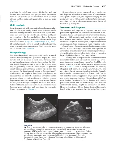
FIG 37.5
(A) Typical ultrasonographic appearance of acute pancreatitis in a Miniature Schnauzer
with a diffusely hypoechoic pancreas (gray arrows) with surrounding hyperechoic
mesentery. (B) Typical ultrasonographic appearance of chronic pancreatitis in an English
Cocker Spaniel. There is a mass-like effect displacing the duodenum. Many dogs and cats
with chronic pancreatitis have an unremarkable abdominal ultrasound. (Courtesy
Diagnostic Imaging Department, Queen’s Veterinary School Hospital, University of
Cambridge, Cambridge, England.)