
Page 654 - Small Animal Internal Medicine, 6th Edition
P. 654
626 PART IV Hepatobiliary and Exocrine Pancreatic Disorders
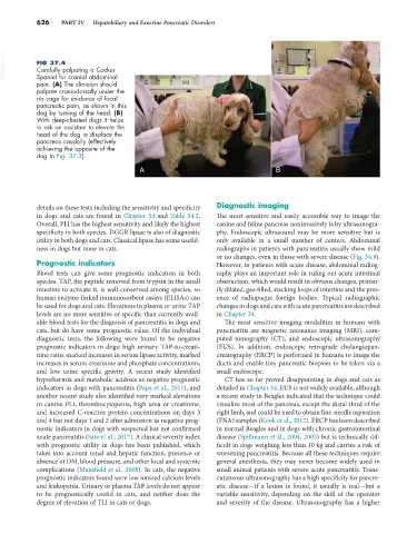
VetBooks.ir FIG 37.4
Carefully palpating a Cocker
Spaniel for cranial abdominal
pain. (A) The clinician should
palpate craniodorsally under the
rib cage for evidence of focal
pancreatic pain, as shown in this
dog by turning of the head. (B)
With deep-chested dogs it helps
to ask an assistant to elevate the
head of the dog to displace the
pancreas caudally (effectively
achieving the opposite of the
dog in Fig. 37.3).
A B
details on these tests including the sensitivity and specificity Diagnostic imaging
in dogs and cats are found in Chapter 34 and Table 34.2. The most sensitive and easily accessible way to image the
Overall, PLI has the highest sensitivity and likely the highest canine and feline pancreas noninvasively is by ultrasonogra-
specificity in both species. DGGR lipase is also of diagnostic phy. Endoscopic ultrasound may be more sensitive but is
utility in both dogs and cats. Classical lipase has some useful- only available in a small number of centers. Abdominal
ness in dogs but none in cats. radiographs in patients with pancreatitis usually show mild
or no changes, even in those with severe disease (Fig. 34.8).
Prognostic indicators However, in patients with acute disease, abdominal radiog-
Blood tests can give some prognostic indication in both raphy plays an important role in ruling out acute intestinal
species. TAP, the peptide removed from trypsin in the small obstruction, which would result in obvious changes, primar-
intestine to activate it, is well conserved among species, so ily dilated, gas-filled, stacking loops of intestine and the pres-
human enzyme-linked immunosorbent assays (ELISAs) can ence of radiopaque foreign bodies. Typical radiographic
be used for dogs and cats. Elevations in plasma or urine TAP changes in dogs and cats with acute pancreatitis are described
levels are no more sensitive or specific than currently avail- in Chapter 34.
able blood tests for the diagnosis of pancreatitis in dogs and The most sensitive imaging modalities in humans with
cats, but do have some prognostic value. Of the individual pancreatitis are magnetic resonance imaging (MRI), com-
diagnostic tests, the following were found to be negative puted tomography (CT), and endoscopic ultrasonography
prognostic indicators in dogs: high urinary TAP-to-creati- (EUS). In addition, endoscopic retrograde cholangiopan-
nine ratio, marked increases in serum lipase activity, marked creatography (ERCP) is performed in humans to image the
increases in serum creatinine and phosphate concentrations, ducts and enable tiny pancreatic biopsies to be taken via a
and low urine specific gravity. A recent study identified small endoscope.
hypothermia and metabolic acidosis as negative prognostic CT has so far proved disappointing in dogs and cats as
indicators in dogs with pancreatitis (Pápa et al., 2011), and detailed in Chapter 34. EUS is not widely available, although
another recent study also identified very marked elevations a recent study in Beagles indicated that the technique could
in canine PLI, thrombocytopenia, high urea or creatinine, visualize most of the pancreas, except the distal third of the
and increased C-reactive protein concentrations on days 3 right limb, and could be used to obtain fine-needle aspiration
and 4 but not days 1 and 2 after admission as negative prog- (FNA) samples (Kook et al., 2012). ERCP has been described
nostic indicators in dogs with suspected but not confirmed in normal Beagles and in dogs with chronic gastrointestinal
acute pancreatitis (Sato et al., 2017). A clinical severity index disease (Spillmann et al., 2004, 2005) but is technically dif-
with prognostic utility in dogs has been published, which ficult in dogs weighing less than 10 kg and carries a risk of
takes into account renal and hepatic function, presence or worsening pancreatitis. Because all these techniques require
absence of DM, blood pressure, and other local and systemic general anesthesia, they may never become widely used in
complications (Mansfield et al., 2008). In cats, the negative small animal patients with severe acute pancreatitis. Trans-
prognostic indicators found were low ionized calcium levels cutaneous ultrasonography has a high specificity for pancre-
and leukopenia. Urinary or plasma TAP levels do not appear atic disease—if a lesion is found, it usually is real—but a
to be prognostically useful in cats, and neither does the variable sensitivity, depending on the skill of the operator
degree of elevation of TLI in cats or dogs. and severity of the disease. Ultrasonography has a higher