
Page 687 - Small Animal Internal Medicine, 6th Edition
P. 687
CHAPTER 39 Diagnostic Tests for the Urinary System 659
considerable interassay variation in SCr concentration mea- often follows fluid therapy and reflects decreased tubular
surements. As a consequence, comparing an individual ani- reabsorption of urea rather than an increased GFR.
VetBooks.ir mal’s fasting SCr concentration to previous results from the SYMMETRIC DIMETHYLARGININE
same animal over time (so-called trending) rather than to a
laboratory reference range can improve the clinician’s ability
posttranslational methylation of arginine residues in pro-
to identify progressive kidney disease and establish a SDMA is a low-molecular-weight (113 Da) by-product of
prognosis. teins. More than 90% of SDMA is removed from the body
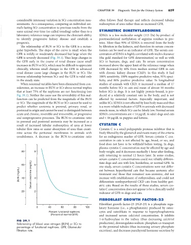
The relationship of BUN or SCr to the GFR is a rectan- by filtration in the kidneys, and therefore its serum concen-
gular hyperbola. The slope of the curve is small when the tration can be used as an indicator of GFR. The serum con-
GFR is mildly or moderately decreased but large when the centration of SDMA is highly correlated with inulin clearance
GFR is severely decreased (Fig. 39.1). Thus large changes in (the gold standard for GFR determination) as well as with
the GFR early in the course of renal disease cause small SCr in humans, dogs, and cats. Its serum concentration
increases in BUN or SCr, which may be difficult to appreciate increased above the upper limit of the reference range when
clinically, whereas small changes in the GFR in advanced GFR decreased by 40% from median normal GFR in cats
renal disease cause large changes in the BUN or SCr. The with chronic kidney disease (CKD). In this study, it had
inverse relationship between SCr and the GFR is valid only 100% sensitivity, 100% negative predictive value, 91% speci-
in the steady state. ficity, and 86% positive predictive value. In longitudinal
When nonrenal variables have been eliminated from con- studies of animals with CKD, it increased a median of 17
sideration, an increase in BUN or SCr above normal implies months before SCr in cats and mean of almost 10 months
that at least 75% of the nephrons are not functioning (see before SCr in dogs. It is not highly protein-bound, is pro-
Fig. 39.1). Neither the cause nor the reversibility of this mal- duced at a relatively constant rate in the body, and is not
function can be predicted from the magnitude of the BUN affected by nonrenal factors. Especially important, and
or SCr. The magnitude of the BUN or SCr cannot be used to unlike SCr, SDMA is not affected by lean body mass and thus
predict whether azotemia is prerenal, primary renal, or is a more reliable indicator of GFR in animals with decreased
postrenal in origin and cannot be used to distinguish between muscle mass, in which SCr can be falsely low. Normal serum
acute and chronic, reversible and irreversible, or progressive SDMA concentrations are < 14 µg/dL in adult dogs and cats
and nonprogressive processes. The BUN-to-creatinine ratio and < 16 µg/dL in puppies and kittens.
in prerenal and postrenal azotemia may be increased as a
result of increased tubular reabsorption of urea at lower CYSTATIN C
tubular flow rates or easier absorption of urea than creati- Cystatin C is a small polypeptide protease inhibitor that is
nine across the peritoneal membranes in animals with freely filtered by the glomeruli and meets many of the criteria
uroabdomen. A decrease in the BUN-to-creatinine ratio for an endogenous marker of GFR. Serum cystatin C con-
centration in cats is not affected by age, breed, or sex, and
food does not have to be withheld before testing. In dogs,
plasma cystatin C concentration may be affected by age and
80 8 body weight, and it decreases markedly 1 hour after feeding,
only returning to normal 12 hours later. In some studies,
serum cystatin C concentrations could not reliably differen-
60 6 tiate dogs and cats with low, borderline, or normal GFR. In
one study, serum cystatin C concentrations were not differ-
BUN (mg/dL) 40 Serum creatinine (mg/dL) 4 ent between hyperthyroid cats that became azotemic after
treatment and those that remained non-azotemic, did not
increase with establishment of euthyroidism, and could not
differentiate nonhyperthyroid CKD cats from healthy geri-
tatin C concentration does not appear to be a clinically useful
20 2 atric cats. Based on the results of these studies, serum cys-
indicator of GFR in dogs and cats.
FIBROBLAST GROWTH FACTOR-23
Fibroblast growth factor-23 (FGF-23) is a phosphate regu-
0 40 60 80 100 latory hormone (i.e., a phosphatonin) produced by osteo-
Percent functional nephrons cytes and osteoblasts in response to hyperphosphatemia
(Percent of normal GFR) and increased serum calcitriol concentration. It inhibits
1-α-hydroxylase in the kidney (thus decreasing calcitriol
FIG 39.1
Relationship of blood urea nitrogen (BUN) or SCr to production), downregulates sodium-phosphate co-transport
percentage of functional nephrons. GFR, Glomerular in the proximal tubules (thus increasing urinary phosphate
filtration rate. excretion), and decreases parathyroid hormone secretion by