
Page 683 - Small Animal Internal Medicine, 6th Edition
P. 683
CHAPTER 38 Clinical Manifestations of Urinary Disorders 655
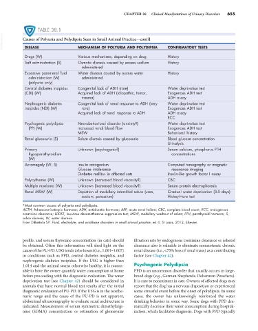
TABLE 38.1
VetBooks.ir Causes of Polyuria and Polydipsia Seen in Small Animal Practice—cont’d CONFIRMATORY TESTS
DISEASE
MECHANISM OF POLYURIA AND POLYDIPSIA
Drugs (W) Various mechanisms, depending on drug History
Salt administration (S) Osmotic diuresis caused by excess sodium History
administered
Excessive parenteral fluid Water diuresis caused by excess water History
administration (W) administered
(polyuria only)
Central diabetes insipidus Congenital lack of ADH (rare) Water deprivation test
(CDI) (W) Acquired lack of ADH (idiopathic, tumor, Exogenous ADH test
trauma) ADH assay
Nephrogenic diabetes Congenital lack of renal response to ADH (very Water deprivation test
insipidus (NDI) (W) rare) Exogenous ADH test
Acquired lack of renal response to ADH ADH assay
ECC
Psychogenic polydipsia Neurobehavioral disorder (anxiety?) Water deprivation test
(PP) (W) Increased renal blood flow Exogenous ADH test
MSW Behavioral history
Renal glucosuria (S) Solute diuresis caused by glucosuria Blood glucose concentration
Urinalysis
Primary Unknown (psychogenic?) Serum calcium, phosphorus PTH
hypoparathyroidism concentrations
(W)
Acromegaly (W, S) Insulin antagonism Computed tomography or magnetic
Glucose intolerance resonance imaging
Diabetes mellitus in affected cats Insulin-like growth factor I assay
Polycythemia (W) Unknown (increased blood viscosity?) CBC
Multiple myeloma (W) Unknown (increased blood viscosity?) Serum protein electrophoresis
Renal MSW (W) Depletion of medullary interstitial solute (urea, Gradual water deprivation (3-5 days)
sodium, potassium) Hickey-Hare test
*Most common causes of polyuria and polydipsia.
ACTH, Adrenocorticotropic hormone; ADH, antidiuretic hormone; ARF, acute renal failure; CBC, complete blood count; ECC, endogenous
creatinine clearance; LDDST, low-dose dexamethasone suppression test; MSW, medullary washout of solute; PTH, parathyroid hormone; S,
solute diuresis; W, water diuresis.
From DiBartola SP: Fluid, electrolyte, and acid-base disorders in small animal practice, ed 4, St Louis, 2012, Elsevier.
profile, and serum thyroxine concentration (in cats) should filtration rate by endogenous creatinine clearance or iohexol
be obtained. Often this information will shed light on the clearance also is valuable to eliminate nonazotemic chronic
cause of the PU-PD. USG tends to be lowest (i.e., 1.001–1.007) renal disease (i.e., <75% loss of renal mass) as a contributing
in conditions such as PPD, central diabetes insipidus, and factor (see Chapter 42).
nephrogenic diabetes insipidus. If the USG is higher than
1.014 and the animal seems otherwise healthy, it is reason- Psychogenic Polydipsia
able to have the owner quantify water consumption at home PPD is an uncommon disorder that usually occurs in large-
before proceeding with the diagnostic evaluation. The water breed dogs (e.g., German Shepherds, Doberman Pinschers).
deprivation test (see Chapter 42) should be considered in It is rare to nonexistent in cats. Owners of affected dogs may
animals that have normal blood test results after the initial report that the dog has a nervous disposition or experienced
diagnostic evaluation of PU-PD. If the USG is in the isosthe- some stressful event before the onset of polydipsia. In some
nuric range and the cause of the PU-PD is not apparent, cases, the owner has unknowingly reinforced the water
abdominal ultrasonography to evaluate renal architecture is drinking behavior in some way. Some dogs with PPD dra-
indicated. Measurement of serum symmetric dimethylargi- matically decrease their water consumption during hospital-
nine (SDMA) concentration or estimation of glomerular ization, which facilitates diagnosis. Dogs with PPD typically