
Page 678 - Small Animal Internal Medicine, 6th Edition
P. 678
650 PART V Urinary Tract Disorders
that may accompany nephrotic syndrome (e.g., glomerular PRESENTING PROBLEMS
disease). Examine the oral cavity for ulcers, tongue tip necro- HEMATURIA
VetBooks.ir sis, and pallor of the mucous membranes. Note retinal Hematuria can be caused by any disease that compromises
edema, detachment, hemorrhage, or vascular tortuosity
during the fundic examination. Occasionally severe hyper-
be associated with diseases of the urinary tract (i.e., kidneys,
tension secondary to renal disease will result in acute onset the urogenital mucosa and results in bleeding. Thus it may
of blindness caused by retinal detachment. Young growing ureters, bladder, urethra) or genital tract (i.e., prostate, penis,
animals with renal failure may develop marked fibrous prepuce, uterus, vagina, vestibule). Hematuria may be clas-
osteodystrophy characterized by enlargement and deformity sified as macroscopic (i.e., visible to the naked eye) or micro-
of the maxilla and mandible (so-called rubber jaw), but this scopic (i.e., identified only as increased numbers of red blood
is rare in older dogs with renal failure. cells in the urine sediment). Macroscopic hematuria results
Both kidneys can be palpated in most cats and the left in a red, pink, or brown coloration of the urine. Centrifuga-
kidney in some dogs. Kidneys should be evaluated for size, tion of the urine sample readily allows differentiation of
shape, consistency, pain, and location. Unless empty, the pigmenturia (e.g., hemoglobinuria, myoglobinuria) from
bladder can be palpated in most dogs and cats. The bladder hematuria (i.e., a pellet of red cells with clear yellow super-
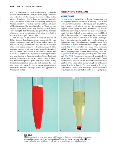
should be evaluated for degree of distention, pain, wall thick- natant; Fig. 38.1). Disorders associated with hematuria
ness, and presence of intramural (e.g., tumors) or intralumi- include urinary tract infection, neoplasia, urolithiasis,
nal (e.g., calculi, clots) masses. In the absence of obstruction, trauma, coagulopathies, vascular anomalies (e.g., renal tel-
a distended bladder in a dehydrated animal suggests abnor- angiectasia in Welsh Corgi dogs), and idiopathic renal hema-
mal renal function or the administration of drugs that impair turia (Box 38.1). Cystocentesis is commonly associated with
urinary concentrating ability (e.g., glucocorticoids, diuret- microscopic hematuria, and a voided sample should always
ics). Evaluate the prostate gland and pelvic urethra during be obtained to evaluate for this possibility when abnormal
the rectal examination. Exteriorize and examine the penis, numbers of red blood cells (e.g., >three/high-power field) are
and palpate the testes. Perform a vaginal examination to observed in the sediment of a urine sample collected by
evaluate for abnormal discharge, masses, and appearance of cystocentesis. Occasionally microscopic hematuria caused
the urethral orifice. by cystocentesis is interpreted as evidence of ongoing feline
A B
FIG 38.1
(A) Unspun urine sample from a dog with hematuria. Without centrifugation, one cannot
differentiate pigmenturia (e.g., hemoglobinuria) from hematuria (i.e., red cells). (B) Red
blood cell pellet after centrifugation of a urine sample from a dog with hematuria.