
Page 705 - Small Animal Internal Medicine, 6th Edition
P. 705
CHAPTER 40 Glomerular Disease 677
circulating immune complexes may become trapped in MECHANISMS OF IMMUNE INJURY
the glomeruli in conditions of antigen-antibody equiva-
VetBooks.ir lence or slight antigen excess. Immune complexes also may Immune complex deposition in glomeruli may decrease the
amount of fixed negative charge and increase the filtration
be formed in situ in response to endogenous glomerular
of negatively charged circulating macromolecules (e.g.,
antigens, endogenous nonglomerular antigens, or exog-
enous antigens deposited or planted in the glomerular filter. albumin). Complement activation results in membrane
Immune complexes may be deposited in subepithelial, sub- damage and proteinuria, and soluble complement compo-
endothelial, or intramembranous locations in the glomerular nents recruit inflammatory cells. Platelet activation and
capillary wall or in the mesangium. Factors affecting the aggregation may occur because of endothelial damage or
location of these deposits include the size and charge of the antigen–antibody interaction, thus exacerbating glomerular
complexes as well as the potential removal of complexes damage by release of a variety of mediators. These mediators
by phagocytosis. The location of deposition contributes cause activation and proliferation of mesangial cells and
to the histopathologic findings and severity of glomerular endothelial cells, vasospasm, and local hypercoagulability.
dysfunction. Neutrophils and macrophages localize in the glomeruli in
Immune complexes can be detected in the glomeruli response to soluble mediators, including complement com-
by staining renal tissue sections with fluorescein-labeled ponents, platelet activating factor, platelet–derived growth
antibody against immunoglobulins or complement of the factor, and eicosanoids. Activated neutrophils release reac-
species being studied. This technique requires renal biopsy tive oxygen species and proteinases, leading to additional
specimens to be collected and sent to the diagnostic labora- damage. Macrophages produce proteinases, oxidants, eico-
tory in special preservative solutions (e.g., Michel’s solution). sanoids, growth factors, cytokines, complement fragments,
More recently, immunohistochemistry using peroxidase- and coagulation factors. Several infectious and inflammatory
antiperoxidase methods have been applied to specimens diseases have been associated with glomerular deposition or
preserved routinely in 10% buffered formalin. Glomerular in situ formation of immune complexes in dogs and cats
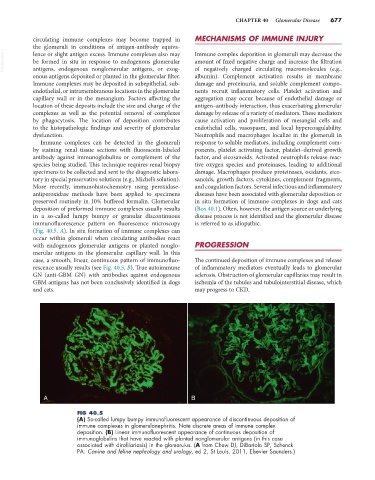
deposition of preformed immune complexes usually results (Box 40.1). Often, however, the antigen source or underlying
in a so-called lumpy bumpy or granular discontinuous disease process is not identified and the glomerular disease
immunofluorescence pattern on fluorescence microscopy is referred to as idiopathic.
(Fig. 40.5, A). In situ formation of immune complexes can
occur within glomeruli when circulating antibodies react
with endogenous glomerular antigens or planted nonglo- PROGRESSION
merular antigens in the glomerular capillary wall. In this
case, a smooth, linear, continuous pattern of immunofluo- The continued deposition of immune complexes and release
rescence usually results (see Fig. 40.5, B). True autoimmune of inflammatory mediators eventually leads to glomerular
GN (anti-GBM GN) with antibodies against endogenous sclerosis. Obstruction of glomerular capillaries may result in
GBM antigens has not been conclusively identified in dogs ischemia of the tubules and tubulointerstitial disease, which
and cats. may progress to CKD.
A B
FIG 40.5
(A) So-called lumpy bumpy immunofluorescent appearance of discontinuous deposition of
immune complexes in glomerulonephritis. Note discrete areas of immune complex
deposition. (B) Linear immunofluorescent appearance of continuous deposition of
immunoglobulins that have reacted with planted nonglomerular antigens (in this case
associated with dirofilariasis) in the glomerulus. (A from Chew DJ, DiBartola SP, Schenck
PA: Canine and feline nephrology and urology, ed 2, St Louis, 2011, Elsevier Saunders.)