
Page 707 - Small Animal Internal Medicine, 6th Edition
P. 707
CHAPTER 40 Glomerular Disease 679
fusion can be detected by electron microscopy. Ultrastruc- reactive, immunoglobulin-associated, and heredofamilial
tural changes include thickening or splitting of the basement syndromes.
VetBooks.ir membrane, podocyte foot process fusion, increased cellular- characterized by tissue deposition of amyloid A protein (AA
Reactive (secondary) amyloidosis is a systemic syndrome
ity of the mesangial space, and the presence of electron-
amyloid). Naturally occurring systemic amyloidosis in
dense deposits (i.e., immune complexes).
domestic animals is an example of reactive amyloidosis.
Familial amyloid syndromes in the Abyssinian, Siamese, and
AMYLOIDOSIS Oriental Shorthair breeds of cat and in the Shar Pei, Beagle,
and English Foxhound breeds of dog are examples of reactive
Amyloidosis refers to a diverse group of diseases character- systemic amyloidosis.
ized by extracellular deposition of fibrils formed by poly- Tissue deposits in animals with reactive systemic amyloi-
merization of protein subunits, with a specific biophysical dosis contain amyloid A protein, which is an amino terminal
conformation called the β-pleated sheet. This specific bio- fragment of an acute-phase reactant called serum amyloid A
physical conformation is responsible for the unique optical protein (SAA). Serum amyloid A protein is one of several
and tinctorial properties of amyloid deposits as well as their acute-phase reactants synthesized by the liver in response to
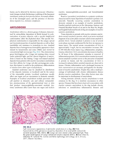
insolubility and resistance to proteolysis in vivo. Amyloid tissue injury. The normal serum concentration of SAA is
deposits have a homogeneous eosinophilic appearance when approximately 1 mg/L, but its concentration increases 100-
stained by hematoxylin and eosin (H&E) and viewed by to 500-fold after tissue injury (e.g., inflammation, neoplasia,
conventional light microscopy (Fig. 40.6). They demonstrate trauma, infarction). SAA concentration decreases to baseline
green birefringence after Congo red staining when viewed by 48 hours if the inflammatory stimulus is removed. If
under polarized light, and the clinical diagnosis of amyloi- inflammation persists, SAA concentration remains increased.
dosis is based on this finding. Congo red-stained amyloid Serum amyloid A protein serves as the precursor of amyloid
deposits from patients with reactive (secondary) amyloidosis A protein in tissues, and the concentration of SAA is
lose their affinity for Congo red after permanganate oxida- increased in plasma before amyloid deposits are observed in
tion; this feature is useful in the preliminary differentiation tissues. Chronic inflammation and a prolonged increase in
of reactive from other types of amyloidosis. SAA concentration are necessary prerequisites for develop-
Amyloid syndromes may be classified by distribution ment of reactive amyloidosis. Despite this, only a small per-
of the deposits (systemic or localized) and by the nature centage of individuals with chronic inflammatory disease
of the responsible protein. Localized syndromes usually develop reactive amyloidosis. Thus other factors must also
affect one organ and are uncommon in domestic animals. be important in development of amyloidosis.
Examples of localized amyloidosis include pancreatic islet Among domestic animals, reactive amyloidosis is most
cell amyloid in domestic cats and solitary extramedul- common in the dog. It is relatively uncommon in other
lary plasmacytomas of the gastrointestinal tract or skin species. Diseases that have been observed in association with
that produce immunoglobulin-associated amyloid. Sys- reactive systemic amyloidosis in the dog include chronic
temic syndromes affect more than one organ and include infectious or noninfectious inflammatory diseases and
A B
FIG 40.6
Light microscopic appearance of glomerular amyloidosis in a dog. (A) Note
hypocellularity of the glomerulus caused by deposition of an eosinophilic extracellular
material (amyloid; H&E, 400×). (B) Congo red stain viewed under polarized light. Note
the green birefringence of Congo red-stained amyloid deposits (400×). (A from Chew DJ,
DiBartola SP, Schenck PA: Canine and feline nephrology and urology, ed 2, St Louis,
2011, Elsevier Saunders.)