
Page 718 - Small Animal Internal Medicine, 6th Edition
P. 718
690 PART V Urinary Tract Disorders
The kidneys are normal-sized or enlarged and of normal of supportive care to determine whether adequate renal
shape in patients with AKI. Renal ultrasonography may show function is likely to return. The severity of residual azotemia
VetBooks.ir increased cortical or medullary echogenicity, but normal fully as a CKD patient.
will determine whether the animal can be managed success-
ultrasonographic examination findings do not exclude AKI.
Initially, the most life-threatening disturbances should be
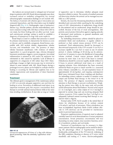
The kidneys of animals with ethylene glycol intoxication are
extremely hyperechoic, and this observation may be helpful identified and corrected while searching for the underlying
diagnostically (Fig. 41.4). Radiographic signs of pulmonary cause of AKI. Administration of nephrotoxic drugs should
disease (e.g., alveolar pattern, alveolar mineralization) were be discontinued and no nephrotoxic drugs should be pre-
more common in dogs with AKI than in those with CKD in scribed. Because of the loss of renal autoregulation, AKI
one study, but these findings did not affect survival. Acute patients cannot protect themselves against ongoing episodes
and convalescent serology testing is useful to establish a of decreased renal perfusion, so general anesthesia and
diagnosis of leptospirosis in dogs with acute nephritis. surgery should be avoided.
Renal biopsy is used to confirm that azotemia is caused An indwelling intravenous catheter should be placed to
by primary renal lesions, to characterize the lesions as acute administer fluids and medications. A jugular catheter is
or chronic, and to establish a prognosis. Renal lesions com- preferred so that central venous pressure (CVP) can be
patible with AKI include tubular degeneration, tubular monitored. Fluid administration should be decreased or
necrosis, and intratubular casts. The presence of intact discontinued temporarily if the CVP exceeds 13 cm H 2 O or
tubular basement membranes with evidence of tubular increases rapidly by 2 cm H 2 O or more in any 10-minute
regeneration is a good prognostic sign, whereas disrupted period. A volume challenge of 20 mL/kg can be adminis-
basement membranes suggest a worse prognosis. Interstitial tered over 10 minutes to assess the likelihood of impending
inflammation is minimal in AKI caused by nephrosis but volume overload. Central venous pressure should not
substantial in AKI caused by nephritis. Lack of fibrosis is increase by more than 2 cm H 2 O if the heart is normal.
supportive of a diagnosis of AKI rather than CKD. Histo- Dehydration should be corrected rapidly, ideally within 6 to
pathologic changes on light microscopy may be minimal to 8 hours to prevent additional renal injury as a result of
absent in some animals with AKI. Renal biopsy during a ongoing ischemia. Once dehydration has been corrected,
prolonged recovery phase can be helpful to evaluate whether additional fluids are given to match sensible (i.e., measured
healing is occurring by fibrosis and nephron loss or by urine volume), insensible (i.e., GI and respiratory losses of
tubular regeneration and repopulation of intact basement approximately 20 mL/kg/day), and ongoing contemporary
membranes. fluid losses (estimated losses from vomiting and diarrhea).
An indwelling urinary catheter is needed to monitor urine
Treatment output and facilitate fluid therapy in the initial 24 to 48
The ultimate goal in management of the maintenance phase hours. The presence of oliguria necessitates meticulous
of AKI is to provide adequate supportive care and time for attention to fluid therapy to prevent overhydration. Weigh-
healing to occur. Prevention of additional renal injury is an ing the patient twice daily on the same scale will provide
important treatment goal; this requires conscientious fluid useful information about fluid balance. Normal urine output
therapy to provide optimal renal perfusion while at the same is 1 to 2 mL/kg/h, and a urine output of 2 to 5 mL/kg/h is
time avoiding overhydration. It may take as long as 3 weeks expected in normal dogs and cats receiving adequate fluid
volume expansion. Urine output less than 2 mL/kg/h in an
adequately hydrated animal receiving fluid therapy is con-
sidered relative oliguria.
Normal saline (0.9% NaCl) usually is the initial fluid of
choice for rehydration because of its sodium content
(154 mEq/L) and lack of potassium. When rehydration has
been accomplished, hypotonic fluids (0.45% sodium chlo-
ride in 2.5% dextrose) can be provided for maintenance
needs to prevent hypernatremia.
Potassium supplementation, if required, must be adjusted
carefully based on serial determinations of serum potassium
concentration. Serum potassium concentration will vary
depending on urine output, renal excretory function, sever-
ity of metabolic acidosis, and oral intake.
Treatment of hyperkalemia may be necessary in oligo-
anuric patients. Electrocardiography can be useful for detect-
ing the physiologic effects of hyperkalemia, including
FIG 41.4
Ultrasound appearance of kidney of a dog with ethylene bradycardia, prolongation of the P-R interval, widening of
glycol poisoning. Note the extremely hyperechoic renal the QRS complexes, blunting or absence of P waves (atrial
cortex. standstill), and tenting of T waves. Electrocardiographic