
Page 754 - Small Animal Internal Medicine, 6th Edition
P. 754
726 PART V Urinary Tract Disorders
diagnostic tests such as contrast cystourethrography, abdom- other electrolyte and acid–base disturbances such as hypo-
inal ultrasonography, and even cystoscopy (Figs. 44.2 and calcemia and acidosis. If hyperkalemia is present, IV treat-
VetBooks.ir 44.3; Video 44.1, cystoscopy of FIC cat) can be performed in ment with fluids, regular insulin (0.25-0.5 U/kg, slow bolus),
and 50% dextrose should be administered. An electrocardio-
recurrent cases to be certain no other disease could account
gram should be evaluated and, in more severe cases, 10% IV
for the present clinical signs.
calcium gluconate may be warranted to counteract the effects
of the hyperkalemia on cardiac conduction. Acidosis is
TREATMENT OPTIONS usually corrected with fluid therapy, but IV sodium bicar-
bonate (1-2 mEq/kg) can also be considered for cats with
ACUTE EPISODES severe hyperkalemia. Care should be taken with bicarbonate
Obstructive Feline Idiopathic Cystitis infusions because exacerbation of hypocalcemia can occur
Once the diagnosis of a urethral obstruction is made, the cat when the acidosis is corrected.
should be assessed and stabilized with intravenous (IV) Once the cat is stabilized, an abdominal radiograph
fluids. A serum biochemical panel should be submitted to should be obtained to evaluate for the most common calculi
evaluate for postrenal azotemia, possible hyperkalemia, and reported in cats (struvite and calcium oxalate [CaOx]). To
provide an immediate reservoir for urine flow, a decompres-
sive cystocentesis should then be performed. Usually a
22-gauge 1- or 1.5-inch needle is inserted into the bladder,
0 with the bevel aimed at the trigone. The needle is connected
to an extension set, three-way stopcock, and 20- or 35-mL
1 syringe (Fig. 44.4). By doing this, all the urine can be drained
without repeated needle insertions into the bladder. Analge-
+ 2 sics should be provided (e.g., buprenorphine, 0.01 mg/kg
SAGITTAL + intravenously q8-12h initially) and once the cat is anesthe-
3 tized (e.g., with isoflurane, sevoflurane, or propofol), the
urethral obstruction can be removed.
4 In rare cases, one can remove a urethral plug by massaging
0.44 cm the distal penis. In most cases, urethral catheterization with
an open-ended nonmetal catheter provides the easiest, safest
means to alleviate the obstruction. The penile urethra should
be clipped and scrubbed; using sterile technique, the catheter
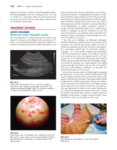
FIG 44.2
Abdominal ultrasonogram from a cat with a urethral is then inserted into the distal urethra. The catheter should
obstruction. The findings are generally nonspecific and also be attached to an extension set and three-way stopcock.
include a thickened bladder wall. This imaging modality is This may help lessen the trauma to the urethra during cath-
not ideal for evaluating the urethra of cats. eter insertion by allowing the assistant more room to flush
the urethra with sterile saline, which is usually required to
dislodge the obstruction; a smaller syringe provides a more
forceful flow, which can be helpful in some cases.
FIG 44.3
Cystoscopic view of a bladder from a female cat with FIC.
The cat has severe edema and increased bladder friability. FIG 44.4
These findings do not appear to correlate well with the Decompressive cystocentesis in a cat with urethral
clinical signs. obstruction.