
Page 759 - Small Animal Internal Medicine, 6th Edition
P. 759
CHAPTER 45 Disorders of Micturition 731
SPINAL CORD L 1-4 S 1-3
VetBooks.ir Kidney Hypogastric Pelvic Pudendal FIG 45.1
nerve
nerve
Ureter β α nerve Schematic diagram of the
sympathetic, parasympathetic, and
somatic innervation to the lower
urinary tract. The micturition pathway
is much more complex than what is
pictured here; the sensory pathways
and higher centers are not depicted
External in this diagram.
urethral
Bladder sphincter
0
1
2
U BLADDER
3
4
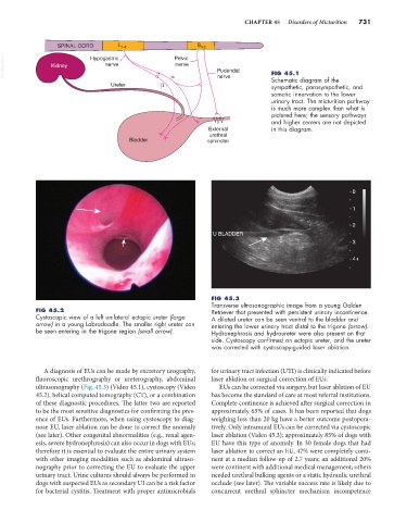
FIG 45.3
Transverse ultrasonographic image from a young Golden
FIG 45.2 Retriever that presented with persistent urinary incontinence.
Cystoscopic view of a left unilateral ectopic ureter (large A dilated ureter can be seen ventral to the bladder and
arrow) in a young Labradoodle. The smaller right ureter can entering the lower urinary tract distal to the trigone (arrow).
be seen entering in the trigone region (small arrow). Hydronephrosis and hydroureter were also present on that
side. Cystoscopy confirmed an ectopic ureter, and the ureter
was corrected with cystoscopy-guided laser ablation.
A diagnosis of EUs can be made by excretory urography, for urinary tract infection (UTI) is clinically indicated before
fluoroscopic urethrography or ureterography, abdominal laser ablation or surgical correction of EUs.
ultrasonography (Fig. 45.3) (Video 45.1), cystoscopy (Video EUs can be corrected via surgery, but laser ablation of EU
45.2), helical computed tomography (CT), or a combination has become the standard of care at most referral institutions.
of these diagnostic procedures. The latter two are reported Complete continence is achieved after surgical correction in
to be the most sensitive diagnostics for confirming the pres- approximately 65% of cases. It has been reported that dogs
ence of EUs. Furthermore, when using cystoscopy to diag- weighing less than 20 kg have a better outcome postopera-
nose EU, laser ablation can be done to correct the anomaly tively. Only intramural EUs can be corrected via cystoscopic
(see later). Other congenital abnormalities (e.g., renal agen- laser ablation (Video 45.3); approximately 85% of dogs with
esis, severe hydronephrosis) can also occur in dogs with EUs; EU have this type of anomaly. In 30 female dogs that had
therefore it is essential to evaluate the entire urinary system laser ablation to correct an EU, 47% were completely conti-
with other imaging modalities such as abdominal ultraso- nent at a median follow-up of 2.7 years; an additional 20%
nography prior to correcting the EU to evaluate the upper were continent with additional medical management; others
urinary tract. Urine cultures should always be performed in needed urethral bulking agents or a static hydraulic urethral
dogs with suspected EUs as secondary UI can be a risk factor occlude (see later). The variable success rate is likely due to
for bacterial cystitis. Treatment with proper antimicrobials concurrent urethral sphincter mechanism incompetence