
Page 793 - Small Animal Internal Medicine, 6th Edition
P. 793
CHAPTER 47 Disorders of the Parathyroid Gland 765
lysis syndrome. (See Chapter 53 for more information on of an abdominal ultrasound. The history and physical exami-
causes of hypocalcemia and hyperphosphatemia in dogs and nation findings are essentially unremarkable in dogs and cats
VetBooks.ir cats.) The diagnosis of primary hypoparathyroidism is estab- with primary hypoparathyroidism, other than those findings
caused by hypocalcemia. The only relevant abnormalities
lished by identifying an undetectable serum PTH concentra-
tion in the face of severe hypocalcemia in a dog or cat in
calcemia and, in most dogs and cats, hyperphosphatemia.
which other causes of hypocalcemia have been ruled out identified on routine blood and urine tests are severe hypo-
(Table 47.3). Most causes of hypocalcemia can be identified Serum total protein, albumin, urea nitrogen, creatinine, and
after evaluation of the history, findings on physical examina- magnesium concentrations are normal. Abdominal ultra-
tion, and results of routine blood and urine tests and review sound is also normal.
Measurement of serum PTH concentration helps confirm
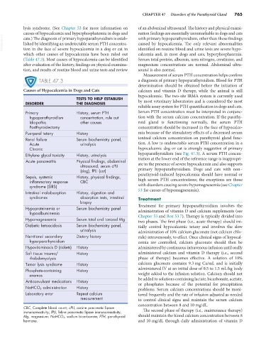
TABLE 47.3 a diagnosis of primary hypoparathyroidism. Blood for PTH
determination should be obtained before the initiation of
Causes of Hypocalcemia in Dogs and Cats calcium and vitamin D therapy, while the animal is still
hypocalcemic. The two-site IRMA system is currently used
TESTS TO HELP ESTABLISH
DISORDER THE DIAGNOSIS by most veterinary laboratories and is considered the most
reliable assay system for PTH quantification in dogs and cats.
Primary History, serum PTH Serum PTH concentration must be interpreted in conjunc-
hypoparathyroidism concentration, rule out tion with the serum calcium concentration. If the parathy-
Idiopathic other causes roid gland is functioning normally, the serum PTH
Postthyroidectomy concentration should be increased in the face of hypocalce-
Puerperal tetany History mia because of the stimulatory effects of a decreased serum
Renal failure Serum biochemistry panel, ionized calcium concentration on parathyroid gland func-
Acute urinalysis tion. A low to undetectable serum PTH concentration in a
Chronic hypocalcemic dog or cat is strongly suggestive of primary
Ethylene glycol toxicity History, urinalysis hypoparathyroidism (see Fig. 47.3). A serum PTH concen-
Acute pancreatitis Physical findings, abdominal tration at the lower end of the reference range is inappropri-
ultrasound, serum cPLI ate in the presence of severe hypocalcemia and also supports
(dog), fPLI (cat) primary hypoparathyroidism. Dogs and cats with non–
Sepsis, systemic History, physical findings, parathyroid-induced hypocalcemia should have normal or
inflammatory response CBC high serum PTH concentrations; the exceptions are those
syndrome (SIRS) with disorders causing severe hypomagnesemia (see Chapter
Intestinal malabsorption History, digestion and 53 for causes of hypomagnesemia).
syndromes absorption tests, intestinal Treatment
biopsy
Hypoproteinemia or Serum biochemistry panel Treatment for primary hypoparathyroidism involves the
hypoalbuminemia administration of vitamin D and calcium supplements (see
Hypomagnesemia Serum total and ionized Mg Chapter 53 and Box 53.7). Therapy is typically divided into
two phases. The first phase (i.e., acute therapy) should ini-
Diabetic ketoacidosis Serum biochemistry panel, tially control hypocalcemic tetany and involves the slow
urinalysis administration of 10% calcium gluconate (not calcium chlo-
Nutritional secondary Dietary history ride) intravenously, to effect. Once clinical signs of hypocal-
hyperparathyroidism cemia are controlled, calcium gluconate should then be
Hypovitaminosis D (rickets) History administered by continuous intravenous infusion until orally
Soft tissue trauma/ History administered calcium and vitamin D therapy (i.e., second
rhabdomyolysis phase of therapy) becomes effective. A solution of 10%
Tumor lysis syndrome History calcium gluconate contains 9.3 mg Ca/mL and is initially
Phosphate-containing History administered IV at an initial dose of 0.5 to 1.5 mL/kg body
enemas weight added to the infusion solution. Calcium should not
Anticonvulsant medications History be added to solutions containing lactate, bicarbonate, acetate,
or phosphates because of the potential for precipitation
NaHCO 3 administration History problems. Serum calcium concentrations should be moni-
Laboratory error Repeat calcium tored frequently and the rate of infusion adjusted as needed
measurement to control clinical signs and maintain the serum calcium
concentration between 8 and 10 mg/dL.
CBC, Complete blood count; cPLI, canine pancreatic lipase
immunoreactivity; fPLI, feline pancreatic lipase immunoreactivity; The second phase of therapy (i.e., maintenance therapy)
Mg, magnesium; NaHCO 3, sodium bicarbonate; PTH, parathyroid should maintain the blood calcium concentration between 8
hormone. and 10 mg/dL through daily administration of vitamin D