Page 788 - Small Animal Internal Medicine, 6th Edition
P. 788
760    PART VI   Endocrine Disorders


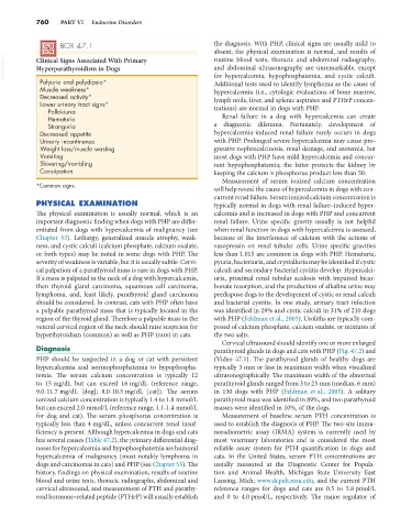
                   BOX 47.1                                      the diagnosis. With PHP, clinical signs are usually mild to
                                                                 absent, the physical examination is normal, and results of
  VetBooks.ir  Clinical Signs Associated With Primary            routine blood tests, thoracic and abdominal radiography,
                                                                 and abdominal ultrasonography are unremarkable, except
            Hyperparathyroidism in Dogs
             Polyuria and polydipsia*                            for hypercalcemia, hypophosphatemia, and cystic calculi.
                                                                 Additional tests used to identify lymphoma as the cause of
             Muscle weakness*                                    hypercalcemia (i.e., cytologic evaluations of bone marrow,
             Decreased activity*                                 lymph node, liver, and splenic aspirates and PTHrP concen-
             Lower urinary tract signs*
               Pollakiuria                                       trations) are normal in dogs with PHP.
               Hematuria                                           Renal failure in a dog with hypercalcemia can create
               Stranguria                                        a diagnostic dilemma. Fortunately, development of
             Decreased appetite                                  hypercalcemia-induced renal failure  rarely occurs in  dogs
             Urinary incontinence                                with PHP. Prolonged severe hypercalcemia may cause pro-
             Weight loss/muscle wasting                          gressive nephrocalcinosis, renal damage, and azotemia, but
             Vomiting                                            most dogs with PHP have mild hypercalcemia and concur-
             Shivering/trembling                                 rent  hypophosphatemia;  the  latter  protects  the  kidney  by
             Constipation                                        keeping the calcium × phosphorus product less than 50.
                                                                   Measurement of serum ionized calcium concentration
            *Common signs.
                                                                 will help reveal the cause of hypercalcemia in dogs with con-
                                                                 current renal failure. Serum ionized calcium concentration is
            PHYSICAL EXAMINATION                                 typically normal in dogs with renal failure–induced hyper-
            The physical examination is usually normal, which is an   calcemia and is increased in dogs with PHP and concurrent
            important diagnostic finding when dogs with PHP are differ-  renal failure. Urine specific gravity usually is not helpful
            entiated from dogs with hypercalcemia of malignancy (see   when renal function in dogs with hypercalcemia is assessed,
            Chapter 53). Lethargy, generalized muscle atrophy, weak-  because of the interference of calcium with the actions of
            ness, and cystic calculi (calcium phosphate, calcium oxalate,   vasopressin on renal tubular cells. Urine specific gravities
            or both types) may be noted in some dogs with PHP. The   less than 1.015 are common in dogs with PHP. Hematuria,
            severity of weakness is variable, but it is usually subtle. Cervi-  pyuria, bacteriuria, and crystalluria may be identified if cystic
            cal palpation of a parathyroid mass is rare in dogs with PHP.   calculi and secondary bacterial cystitis develop. Hypercalci-
            If a mass is palpated in the neck of a dog with hypercalcemia,   uria, proximal renal tubular acidosis with impaired bicar-
            then  thyroid  gland  carcinoma,  squamous  cell  carcinoma,   bonate resorption, and the production of alkaline urine may
            lymphoma,  and,  least  likely,  parathyroid  gland  carcinoma   predispose dogs to the development of cystic or renal calculi
            should be considered. In contrast, cats with PHP often have   and bacterial cystitis. In one study, urinary tract infection
            a palpable parathyroid mass that is typically located in the   was identified in 29% and cystic calculi in 31% of 210 dogs
            region of the thyroid gland. Therefore a palpable mass in the   with PHP (Feldman et al., 2005). Uroliths are typically com-
            ventral cervical region of the neck should raise suspicion for   posed of calcium phosphate, calcium oxalate, or mixtures of
            hyperthyroidism (common) as well as PHP (rare) in cats.  the two salts.
                                                                   Cervical ultrasound should identify one or more enlarged
            Diagnosis                                            parathyroid glands in dogs and cats with PHP (Fig. 47.2) and
            PHP should be suspected in a dog or cat with persistent   (Video 47.1). The parathyroid glands of healthy dogs are
            hypercalcemia and normophosphatemia to hypophospha-  typically 3 mm or less in maximum width when visualized
            temia. The serum calcium concentration is typically 12   ultrasonographically. The maximum width of the abnormal
            to 15 mg/dL but can exceed 16 mg/dL (reference range,   parathyroid glands ranged from 3 to 23 mm (median, 6 mm)
            9.0-11.7 mg/dL  [dog];  8.0-10.5 mg/dL  [cat]).  The  serum   in 130 dogs with PHP (Feldman et al., 2005). A solitary
            ionized calcium concentration is typically 1.4 to 1.8 mmol/L   parathyroid mass was identified in 89%, and two parathyroid
            but can exceed 2.0 mmol/L (reference range, 1.1-1.4 mmol/L   masses were identified in 10%, of the dogs.
            for dog and cat). The serum phosphorus concentration is   Measurement of baseline serum PTH concentration is
            typically less than 4 mg/dL, unless concurrent renal insuf-  used to establish the diagnosis of PHP. The two-site immu-
            ficiency is present. Although hypercalcemia in dogs and cats   noradiometric assay (IRMA) system is currently used by
            has several causes (Table 47.2), the primary differential diag-  most veterinary laboratories and is considered the most
            noses for hypercalcemia and hypophosphatemia are humoral   reliable assay system for PTH quantification in dogs and
            hypercalcemia of malignancy (most notably lymphoma in   cats. In the United States, serum PTH concentrations are
            dogs and carcinomas in cats) and PHP (see Chapter 53). The   usually  measured  at  the  Diagnostic  Center  for  Popula-
            history, findings on physical examination, results of routine   tion and Animal Health, Michigan State University East
            blood and urine tests, thoracic radiographs, abdominal and   Lansing, Mich; www.dcpah.msu.edu, and the current PTH
            cervical ultrasound, and measurement of PTH and parathy-  reference ranges for dogs and cats are 0.5 to 5.8 pmol/L
            roid hormone–related peptide (PTHrP) will usually establish   and 0 to 4.0 pmol/L, respectively. The major regulator of

