
Page 908 - Small Animal Internal Medicine, 6th Edition
P. 908
880 PART VI Endocrine Disorders
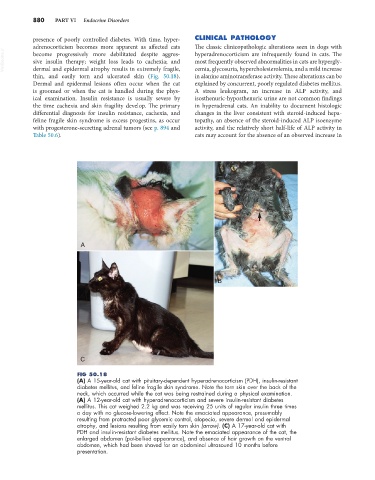
presence of poorly controlled diabetes. With time, hyper- CLINICAL PATHOLOGY
adrenocorticism becomes more apparent as affected cats The classic clinicopathologic alterations seen in dogs with
VetBooks.ir become progressively more debilitated despite aggres- hyperadrenocorticism are infrequently found in cats. The
most frequently observed abnormalities in cats are hypergly-
sive insulin therapy; weight loss leads to cachexia; and
dermal and epidermal atrophy results in extremely fragile,
in alanine aminotransferase activity. These alterations can be
thin, and easily torn and ulcerated skin (Fig. 50.18). cemia, glycosuria, hypercholesterolemia, and a mild increase
Dermal and epidermal lesions often occur when the cat explained by concurrent, poorly regulated diabetes mellitus.
is groomed or when the cat is handled during the phys- A stress leukogram, an increase in ALP activity, and
ical examination. Insulin resistance is usually severe by isosthenuric-hyposthenuric urine are not common findings
the time cachexia and skin fragility develop. The primary in hyperadrenal cats. An inability to document histologic
differential diagnosis for insulin resistance, cachexia, and changes in the liver consistent with steroid-induced hepa-
feline fragile skin syndrome is excess progestins, as occur topathy, an absence of the steroid-induced ALP isoenzyme
with progesterone-secreting adrenal tumors (see p. 894 and activity, and the relatively short half-life of ALP activity in
Table 50.6). cats may account for the absence of an observed increase in
A
B
C
FIG 50.18
(A) A 15-year-old cat with pituitary-dependent hyperadrenocorticism (PDH), insulin-resistant
diabetes mellitus, and feline fragile skin syndrome. Note the torn skin over the back of the
neck, which occurred while the cat was being restrained during a physical examination.
(A) A 12-year-old cat with hyperadrenocorticism and severe insulin-resistant diabetes
mellitus. This cat weighed 2.2 kg and was receiving 25 units of regular insulin three times
a day with no glucose-lowering effect. Note the emaciated appearance, presumably
resulting from protracted poor glycemic control, alopecia, severe dermal and epidermal
atrophy, and lesions resulting from easily torn skin (arrow). (C) A 17-year-old cat with
PDH and insulin-resistant diabetes mellitus. Note the emaciated appearance of the cat, the
enlarged abdomen (pot-bellied appearance), and absence of hair growth on the ventral
abdomen, which had been shaved for an abdominal ultrasound 10 months before
presentation.