
Page 913 - Small Animal Internal Medicine, 6th Edition
P. 913
CHAPTER 50 Disorders of the Adrenal Gland 885
The most challenging aspect of diagnosis is the differen-
BOX 50.9 tiation between acute renal failure and primary adrenal
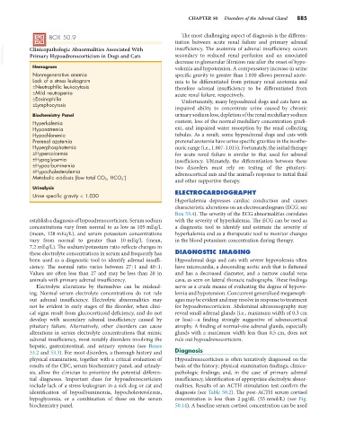
VetBooks.ir Clinicopathologic Abnormalities Associated With insufficiency. The azotemia of adrenal insufficiency occurs
secondary to reduced renal perfusion and an associated
Primary Hypoadrenocorticism in Dogs and Cats
Hemogram decrease in glomerular filtration rate after the onset of hypo-
volemia and hypotension. A compensatory increase in urine
Nonregenerative anemia specific gravity to greater than 1.030 allows prerenal azote-
Lack of a stress leukogram mia to be differentiated from primary renal azotemia and
±Neutrophilic leukocytosis therefore adrenal insufficiency to be differentiated from
±Mild neutropenia acute renal failure, respectively.
±Eosinophilia Unfortunately, many hypoadrenal dogs and cats have an
±Lymphocytosis
impaired ability to concentrate urine caused by chronic
Biochemistry Panel urinary sodium loss, depletion of the renal medullary sodium
Hyperkalemia content, loss of the normal medullary concentration gradi-
Hyponatremia ent, and impaired water resorption by the renal collecting
Hypochloremia tubules. As a result, some hypoadrenal dogs and cats with
Prerenal azotemia prerenal azotemia have urine specific gravities in the isosthe-
Hyperphosphatemia nuric range (i.e., 1.007-1.015). Fortunately, the initial therapy
±Hypercalcemia for acute renal failure is similar to that used for adrenal
±Hypoglycemia insufficiency. Ultimately, the differentiation between these
±Hypoalbuminemia two disorders must rely on testing of the pituitary-
±Hypocholesterolemia adrenocortical axis and the animal’s response to initial fluid
−
Metabolic acidosis (low total CO 2 , HCO 3 )
and other supportive therapy.
Urinalysis
Urine specific gravity < 1.030 ELECTROCARDIOGRAPHY
Hyperkalemia depresses cardiac conduction and causes
characteristic alterations on an electrocardiogram (ECG; see
Box 53.4). The severity of the ECG abnormalities correlates
establish a diagnosis of hypoadrenocorticism. Serum sodium with the severity of hyperkalemia. The ECG can be used as
concentrations vary from normal to as low as 105 mEq/L a diagnostic tool to identify and estimate the severity of
(mean, 128 mEq/L), and serum potassium concentrations hyperkalemia and as a therapeutic tool to monitor changes
vary from normal to greater than 10 mEq/L (mean, in the blood potassium concentration during therapy.
7.2 mEq/L). The sodium/potassium ratio reflects changes in
these electrolyte concentrations in serum and frequently has DIAGNOSTIC IMAGING
been used as a diagnostic tool to identify adrenal insuffi- Hypoadrenal dogs and cats with severe hypovolemia often
ciency. The normal ratio varies between 27 : 1 and 40 : 1. have microcardia, a descending aortic arch that is flattened
Values are often less than 27 and may be less than 20 in and has a decreased diameter, and a narrow caudal vena
animals with primary adrenal insufficiency. cava, as seen on lateral thoracic radiographs. These findings
Electrolyte alterations by themselves can be mislead- serve as a crude means of evaluating the degree of hypovo-
ing. Normal serum electrolyte concentrations do not rule lemia and hypotension. Concurrent generalized megaesoph-
out adrenal insufficiency. Electrolyte abnormalities may agus may be evident and may resolve in response to treatment
not be evident in early stages of the disorder, when clini- for hypoadrenocorticism. Abdominal ultrasonography may
cal signs result from glucocorticoid deficiency, and do not reveal small adrenal glands (i.e., maximum width of 0.3 cm
develop with secondary adrenal insufficiency caused by or less)—a finding strongly suggestive of adrenocortical
pituitary failure. Alternatively, other disorders can cause atrophy. A finding of normal-size adrenal glands, especially
alterations in serum electrolyte concentrations that mimic glands with a maximum width less than 0.5 cm, does not
adrenal insufficiency, most notably disorders involving the rule out hypoadrenocorticism.
hepatic, gastrointestinal, and urinary systems (see Boxes
53.2 and 53.3). For most disorders, a thorough history and Diagnosis
physical examination, together with a critical evaluation of Hypoadrenocorticism is often tentatively diagnosed on the
results of the CBC, serum biochemistry panel, and urinaly- basis of the history; physical examination findings; clinico-
sis, allow the clinician to prioritize the potential differen- pathologic findings; and, in the case of primary adrenal
tial diagnoses. Important clues for hypoadrenocorticism insufficiency, identification of appropriate electrolyte abnor-
include lack of a stress leukogram in a sick dog or cat and malities. Results of an ACTH stimulation test confirm the
identification of hypoalbuminemia, hypocholesterolemia, diagnosis (see Table 50.2). The post-ACTH serum cortisol
hypoglycemia, or a combination of these on the serum concentration is less than 2 µg/dL (55 nmol/L) (see Fig.
biochemistry panel. 50.14). A baseline serum cortisol concentration can be used