
Page 909 - Small Animal Internal Medicine, 6th Edition
P. 909
CHAPTER 50 Disorders of the Adrenal Gland 881
ALP activity. Urine abnormalities frequently identified in interpretation of results (Table 50.3). We rely most heavily
dogs with hyperadrenocorticism are not common in cats. on the UCCR, the dexamethasone suppression test (sensitiv-
VetBooks.ir DIAGNOSTIC IMAGING ity ≈90%), and abdominal ultrasonography to establish the
diagnosis of hyperadrenocorticism in cats. The ACTH stim-
ulation test lacks sensitivity (≈40%) in the cat and is not
Abdominal ultrasonography is used to identify adrenal
masses and to clarify the clinician’s index of suspicion for recommended. We also rely on abdominal ultrasound rather
PDH. Interpretation of results of adrenal imaging in cats is than endogenous plasma ACTH concentration to differenti-
similar to that in dogs (see p. 862). The maximum width of ate PDH from ADH.
the adrenal gland in healthy cats is typically less than 0.5 cm.
Adrenomegaly should be suspected when the maximum Urine Cortisol/Creatinine Ratio
width is greater than 0.5 cm; a maximum width greater than The theory behind and the specifics regarding the UCCR are
0.8 cm is strongly suggestive of adrenomegaly. The finding similar for dogs and cats and are discussed on page 867. The
of easily visualized, bilaterally large adrenals in a cat with UCCR test is a sensitive diagnostic test for identifying hyper-
appropriate clinical signs and physical examination findings adrenocorticism in cats, but similar to dogs, specificity of the
and abnormal test results of the pituitary-adrenocortical axis test is low in cats. We use the UCCR as the initial screening
is strong evidence for PDH. CT and MRI can be used to look test for hyperadrenocorticism in cats. Urine should be col-
for pituitary macroadenoma and to determine the size of an lected at home, preferably on two consecutive days. A normal
adrenal mass and the extent of infiltration of the mass into UCCR in one or both urine samples is strong evidence
surrounding blood vessels and organs before adrenalectomy. against hyperadrenocorticism. An increase in UCCR in both
urine samples does not establish the diagnosis by itself but
TESTS OF THE PITUITARY- supports performing the dexamethasone suppression test.
ADRENOCORTICAL AXIS
Although the tests used to diagnose hyperadrenocorticism Dexamethasone Suppression Test
in cats and dogs are similar (see p. 864), some important The duration of the suppressive effects of intravenously admin-
differences have been noted in the testing protocol and in istered dexamethasone on serum cortisol concentrations is
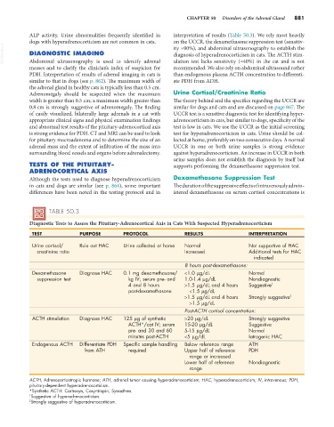
TABLE 50.3
Diagnostic Tests to Assess the Pituitary-Adrenocortical Axis in Cats With Suspected Hyperadrenocorticism
TEST PURPOSE PROTOCOL RESULTS INTERPRETATION
Urine cortisol/ Rule out HAC Urine collected at home Normal Not supportive of HAC
creatinine ratio Increased Additional tests for HAC
indicated
8 hours post-dexamethasone:
Dexamethasone Diagnose HAC 0.1 mg dexamethasone/ <1.0 µg/dL Normal
suppression test kg IV; serum pre- and 1.0-1.4 µg/dL Nondiagnostic
4 and 8 hours >1.5 µg/dL and 4 hours Suggestive †
post-dexamethasone <1.5 µg/dL
>1.5 µg/dL and 4 hours Strongly suggestive ‡
>1.5 µg/dL
Post-ACTH cortisol concentration:
ACTH stimulation Diagnose HAC 125 µg of synthetic >20 µg/dL Strongly suggestive
ACTH*/cat IV; serum 15-20 µg/dL Suggestive
pre- and 30 and 60 5-15 µg/dL Normal
minutes post-ACTH <5 µg/dL Iatrogenic HAC
Endogenous ACTH Differentiate PDH Specific sample handling Below reference range ATH
from ATH required Upper half of reference PDH
range or increased
Lower half of reference Nondiagnostic
range
ACTH, Adrenocorticotropic hormone; ATH, adrenal tumor causing hyperadrenocorticism; HAC, hyperadrenocorticism; IV, intravenous; PDH,
pituitary-dependent hyperadrenocorticism.
*Synthetic ACTH: Cortrosyn, Cosyntropin, Synacthen.
† Suggestive of hyperadrenocorticism.
‡ Strongly suggestive of hyperadrenocorticism.