
Page 130 - Veterinary diagnostic imaging birds exotic pets wildlife
P. 130
126 SECTION I III The Birds
A
B
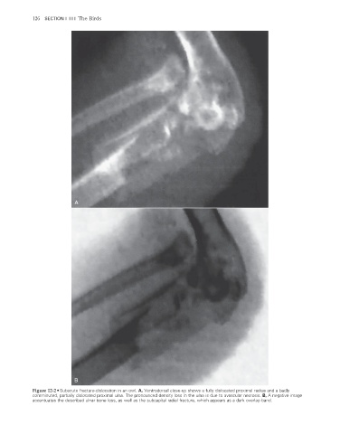
Figure 12-2 • Subacute fracture-dislocation in an owl. A, Ventrodorsal close-up shows a fully dislocated proximal radius and a badly
comminuted, partially dislocated proximal ulna. The pronounced density loss in the ulna is due to avascular necrosis. B, A negative image
accentuates the described ulnar bone loss, as well as the subcapital radial fracture, which appears as a dark overlap band.
2/11/2008 10:57:26 AM
ch012-A02527.indd 126 2/11/2008 10:57:26 AM
ch012-A02527.indd 126