
Page 253 - Veterinary diagnostic imaging birds exotic pets wildlife
P. 253
CHAPTER 21 III Guinea Pigs 249
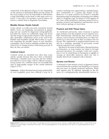
cranial half of the abdomen (Figure 21-19). Depending conduct anything even approaching a standard lame-
on the amount of abdominal detail and the nature of ness examination in a guinea pig (Figure 21-22).
the cecal content, it may be impossible to differentiate However, it is quite common for owners to describe
a large fl uid-filled cecum from a solid mass such as a their guinea pigs as being lame (or limping, not walking
tumor. A mass effect, for example, a gravid uterus, can right, or dragging a leg). So based on what appears to
lead to a similar kind of diagnostic uncertainty. be a clear owner preference, lameness seems to have a
genuine currency and legitimacy and therefore can be
used without apology or reservation.
Bladder Stones (Cystic Calculi)
Cystic calculi, or as they are more commonly termed Fracture and Soft Tissue Injury
bladder stones, are frequently encountered in guinea
pigs and can usually be diagnosed radiographically, As mentioned previously, many fractures in guinea
provided the stones are sufficiently large and dense pigs, perhaps even the majority of such injuries, have
and the image is high quality (Figure 21-20). Occasion- been inadvertently inflicted by their owners. Serious
ally, dystrophic calcification occurs in the wall of the injuries, such as fracture and dislocation, are often sus-
bladder and resembles calculi. Where there is a ques- pected at or near the time of their occurrence but not
tion regarding the possible nature of such densities, always. The undetected or unappreciated fracture can
sonography can be used to establish whether the den- also be a problem, especially in previously owned
sities move or change position while being scanned. If animals acquired from a shelter.
they do, they are stones. But there is far more to limb injury than just fracture
or dislocation, as most readers can attest to through
personal experience. For example, a torn nail, a punc-
Urethral Calculi
ture or cut, or a bad bruise or hematoma is capable of
Urethral calculi are identified less often than cystic causing an observable lameness, albeit one that is
calculi, usually because they have been overlooked. usually short-lived compared with a fracture.
Obviously, a bladder stone can pass into the urethra,
provided it is not too large, with or without accompa- Sprains and Strains
nying urethral colic. Urethral calculi are invariably best
seen in high-quality images of the caudal abdomen A stretched or torn tendon (strain) or ligament (sprain)
(Figure 21-21). can sometimes be as painful and disabling as a fracture
and can cause an equally profound lameness. While
Lameness. At first, the term lameness may seem foolish, such injuries may be inferred, especially in the ab-
or even laughable, given how difficult it may be to sence of a radiographically demonstrable fracture or
Figure 21-19 • Lateral view of the torso of a guinea pig with an impacted cecum shows a caudal abdominal mass or mass effect being
created by organomegaly, most likely the cecum. The unusual appearance of the stomach is the result of cranial displacement by the
enlarged cecum.
2/11/2008 11:09:51 AM
ch021-A02527.indd 249 2/11/2008 11:09:51 AM
ch021-A02527.indd 249