
Page 255 - Veterinary diagnostic imaging birds exotic pets wildlife
P. 255
CHAPTER 21 III Guinea Pigs 251
A
B
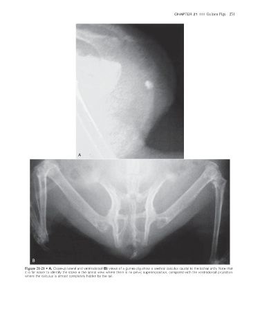
Figure 21-21 • A, Close-up lateral and ventrodorsal (B) views of a guinea pig show a urethral calculus caudal to the ischial arch. Note that
it is far easier to identify the stone in the lateral view where there is no pelvic superimposition, compared with the ventrodorsal projection
where the calculus is almost completely hidden by the tail.
2/11/2008 11:09:52 AM
ch021-A02527.indd 251 2/11/2008 11:09:52 AM
ch021-A02527.indd 251