
Page 306 - Veterinary diagnostic imaging birds exotic pets wildlife
P. 306
302 SECTION II III The Mammals
A B
C D
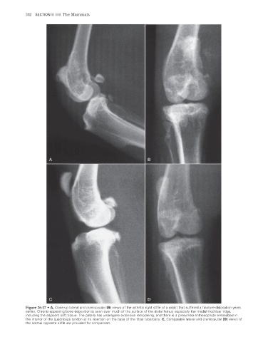
Figure 26-17 • A, Close-up lateral and craniocaudal (B) views of the arthritic right stifle of a rabbit that suffered a fracture-dislocation years
earlier. Chronic-appearing bone deposition is seen over much of the surface of the distal femur, especially the medial trochlear ridge,
including the adjacent soft tissue. The patella has undergone extensive remodeling, and there is a presumed enthesophyte embedded in
the interior of the quadriceps tendon at its insertion on the base of the tibial tuberosity. C, Comparable lateral and craniocaudal (D) views of
the normal opposite stifle are provided for comparison.
2/11/2008 11:12:54 AM
ch026-A02527.indd 302 2/11/2008 11:12:54 AM
ch026-A02527.indd 302