
Page 336 - Veterinary diagnostic imaging birds exotic pets wildlife
P. 336
332 SECTION II III The Mammals
A
B
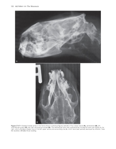
Figure 27-17 • Standard dental series in the ferret shown in previous figures consists of four views: lateral (A), ventrodorsal (B), left
ventrodorsal oblique (C), and right ventrodorsal oblique (D). The ventrodorsal view was customized by moving the lower jaw slightly to the
right, which afforded a clearer view of the left upper canine and surrounding maxilla, which have been partially destroyed by infection. Note
the localized, left-sided facial swelling.
2/11/2008 11:44:09 AM
ch027-A02527.indd 332 2/11/2008 11:44:09 AM
ch027-A02527.indd 332