
Page 340 - Veterinary diagnostic imaging birds exotic pets wildlife
P. 340
336 SECTION II III The Mammals
A
B
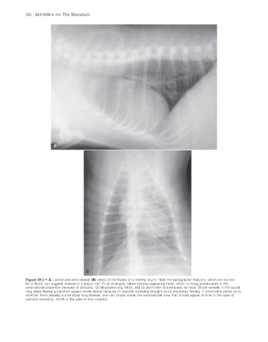
Figure 28-2 • A, Lateral and ventrodorsal (B) views of the thorax of a healthy skunk. Note the radiographic features, which are normal
for a skunk, but suggest disease in a dog or cat: (1) an enlarged, rather bulbous appearing heart, which is being accentuated in the
ventrodorsal projection because of obliquity; (2) fat-grayed lung fields; and (3) prominent extrathoracic fat folds. Blood vessels in the caudal
lung lobes (lateral projection) appear overly dense because of vascular crowding brought on by expiratory filming. If uncertainty exists as to
whether there actually is interstitial lung disease, one can simply check the ventrodorsal view that should appear normal in the case of
vascular crowding, which is the case in this instance.
2/11/2008 11:13:54 AM
ch028-A02527.indd 336 2/11/2008 11:13:54 AM
ch028-A02527.indd 336