
Page 339 - Veterinary diagnostic imaging birds exotic pets wildlife
P. 339
CHAPTER 28 III Skunks, Squirrels, Raccoons, and Armadillos 335
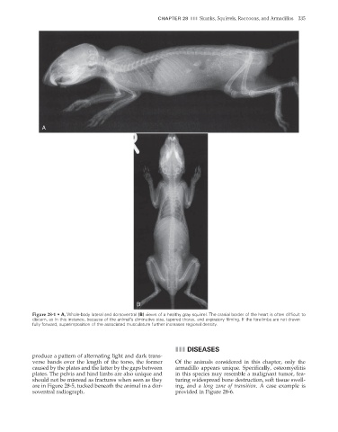
A
B
Figure 28-1 • A, Whole-body lateral and dorsoventral (B) views of a healthy gray squirrel. The cranial border of the heart is often diffi cult to
discern, as in this instance, because of the animal’s diminutive size, tapered thorax, and expiratory filming. If the forelimbs are not drawn
fully forward, superimposition of the associated musculature further increases regional density.
III DISEASES
produce a pattern of alternating light and dark trans-
verse bands over the length of the torso, the former Of the animals considered in this chapter, only the
caused by the plates and the latter by the gaps between armadillo appears unique. Specifi cally, osteomyelitis
plates. The pelvis and hind limbs are also unique and in this species may resemble a malignant tumor, fea-
should not be misread as fractures when seen as they turing widespread bone destruction, soft tissue swell-
are in Figure 28-5, tucked beneath the animal in a dor- ing, and a long zone of transition. A case example is
soventral radiograph. provided in Figure 28-6.
2/11/2008 11:13:53 AM
ch028-A02527.indd 335 2/11/2008 11:13:53 AM
ch028-A02527.indd 335