
Page 361 - Veterinary diagnostic imaging birds exotic pets wildlife
P. 361
CHAPTER 30 III Acreage Pets 357
A B
C
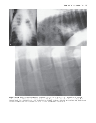
Figure 30-18 • A, Ventrodorsal and lateral (B) views of the chest of a pneumonic miniature goat. Both views are marred by motion
unsharpness because of dyspnea and show bilateral consolidation (“double pneumonia”) and a medium-volume pneumothorax. C, An
ultraclose lateral view of the caudal dorsal thorax reveals the dorsal surface of the partially collapsed right caudal lung lobe, appearing as a
gracefully arched, light gray line crossing the upper half of the image, accentuated by free pleural air.
2/11/2008 11:23:44 AM
ch030-A02527.indd 357 2/11/2008 11:23:44 AM
ch030-A02527.indd 357