
Page 365 - Veterinary diagnostic imaging birds exotic pets wildlife
P. 365
CHAPTER 31 III Performance and Demonstration Pets: Bear and Bison 361
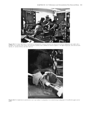
Figure 31-1 • A large black bear is swarmed by radiographers making final positional adjustments before radiographs are made with a
portable x-ray machine (top center). An anesthetic machine and a plethora of monitors appear in the right foreground, while the animal’s
dentist, Dr. James Anthony (far left), looks on.
Figure 31-2 • A black bear is positioned on an x-ray receiver in preparation for a lateral oblique radiograph of the affected upper canine
tooth.
2/11/2008 11:25:49 AM
ch031-A02527.indd 361 2/11/2008 11:25:49 AM
ch031-A02527.indd 361