Page 149 - Withrow and MacEwen's Small Animal Clinical Oncology, 6th Edition
P. 149
128   PART II    Diagnostic Procedures for the Cancer Patient


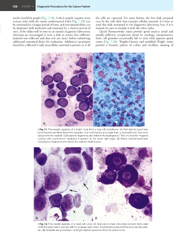
         nuclei should be purple (Fig. 7.1A). A thick sample requires more   the cells are ruptured. For some lesions, the first slide prepared
         contact time with the stains; understained slides (Fig. 7.1B) can   may be the only slide that contains cellular material. It is best to
                                                               send this slide unstained to the diagnostic laboratory, but, if it is
         be restained for a longer period of time, and overstained slides can
  VetBooks.ir  be destained with methanol and restained for a shorter period of   stained, be sure to include it with the other slides.
                                                                  Quick Romanowsky stains provide good nuclear detail and
         time. If the slides will be sent to an outside diagnostic laboratory,
         clinicians are encouraged to stain a slide to ensure that sufficient   usually sufficient cytoplasmic detail for cytologic interpretation.
         material was collected and that cells are intact before submitting   Mast cell granules occasionally fail to stain with aqueous quick
         additional unstained slides for evaluation. Additional specimens   stains (Fig. 7.2A).  Wright–Giemsa and modified  Wright stains
         should be collected if only noncellular material is present or if all   provide a broader palette of colors and excellent staining of


                             A                                 B
                          • Fig. 7.1  Fine-needle aspirate of a lymph node from a dog with lymphoma. (A) Well-stained specimen.
                          Lymphocytes are three times the diameter of an erythrocyte and larger than a neutrophil and have mul-
                          tiple prominent nucleoli. Cytoplasmic fragments are visible in the background. Part of a swollen magenta
                          nucleus with a prominent nucleolus is present at the lower right edge. (B) Poorly stained specimen.
                          Cytoplasmic fragments are visible, but cellular detail is poor.


                        A                                      B
                          • Fig. 7.2  Fine-needle aspirate of a mast cell tumor. (A) Granules in mast cells (large arrows) fail to stain
                          when the specimen is stained with an aqueous quick stain. Small lymphocytes (small arrow) are also pres-
                          ent. (B) Granules are prominent in a Wright-stained specimen from the same tumor.

