Page 150 - Withrow and MacEwen's Small Animal Clinical Oncology, 6th Edition
P. 150
CHAPTER 7  Diagnostic Cytopathology in Clinical Oncology  129


           cytoplasmic granules (Fig. 7.2B), but require more steps and lon-  cytopathologists recognize that primary inflammatory lesions can
           ger staining times or the use of an automated stainer. Fixation of   convincingly mimic neoplastic lesions. (4) If neoplasia is likely,
                                                                 what is the tissue of origin and is the tumor benign or malignant?
           wet smears is required for Papanicolaou staining, which is not fre-
  VetBooks.ir  quently used in veterinary cytology. Heat fixation is not required   These questions can sometimes be answered by cytologic evalua-
                                                                 tion of the lesion but often require confirmation with histologic
           or recommended for cytologic specimens.
             Cytochemical and immunocytochemical staining may be nec-  examination.
           essary to determine the specific tumor type. A complete list of
           available special stains and antibodies is beyond the scope of this   Specimens of Diagnostic Quality
           chapter; consultation with a veterinary cytopathologist is recom-
           mended when considering the necessity and use of these stains.   What constitutes adequate cellularity depends on the type of
           Cytochemical stains identify specific chemical compounds or   tumor. Aspirates of mesenchymal tumors, which often contain
           structures within the cytoplasm or nucleus and include stains such   extracellular matrix, tend to be less cellular than those of epi-
           as Prussian blue for iron; periodic acid–Schiff (PAS) for carbo-  thelial and discrete round cell tumors. The degree of cellular-
                                                       4
           hydrates; alkaline phosphatase for identifying osteoblasts ; and a   ity also has an effect on the level of confidence expressed in the
           wide variety of leukocyte markers, including Sudan black B, per-  interpretation, and diagnostic opinions are often qualified with
           oxidase, chloracetate esterase, and nonspecific esterases. Immu-  “possible” or “probable” for poorly cellular specimens compared
           nocytochemical staining procedures use antibodies to identify   with “diagnostic for” or “consistent with” for highly cellular
           specific proteins or peptides within or on the surface of the cells.   specimens. All cytologic specimens contain some ruptured cells,
           Common antibodies used in veterinary oncology include those   but to render a meaningful interpretation the majority of cells
           directed against CD3 (T cells), CD79a and CD20 (B cells), cyto-  should be intact. Material from ruptured cells is recognized as
           keratin (epithelial cells), vimentin (mesenchymal cells), and Melan   stringy strands of chromatin or swollen magenta nuclei, often
           A (melanocytes). Use of a single stain or antibody is discouraged,   with obvious nucleoli, and free cytoplasmic fragments (see Fig.
           as cell lineage is rarely identified, a single marker is often expressed   7.1A). Large lymphocytes and cells from endocrine tumors are
           by a variety of lineages, and aberrant expression by neoplastic cells   highly susceptible to rupture, and extra care should be taken not
           can occur; a panel of stains or antibodies usually is necessary for   to exert pressure on the cells when preparing cytologic specimens
           complete identification.                              from these lesions.

           Examination and Description of Cytologic              Nonneoplastic Cells and Noncellular Material
           Specimens                                             Found in Cytologic Specimens

           A good microscope, ideally equipped with a digital camera to   The submandibular salivary gland occasionally is aspirated instead
           document cytologic findings for the medical record or for con-  of the mandibular LNs and is recognized by clusters of foamy
           sultation, should be used for examining cytologic specimens. The   cells in a background of mucin and blood. When tissue containing
           4×, 10×, and 20× objectives are useful for scanning the slide and   a metastatic tumor is aspirated, the specimen may contain only
           assessing cellular arrangements and general cell shape, whereas   neoplastic cells or may contain normal cells from the tissue (e.g.,
           the 40× (“high dry”) and 50× or 100× (oil-immersion) objectives   lymphoid populations in an LN), which helps confirm location of
           are required for examining cellular detail. To improve clarity, the   the tumor. Necrosis can be found in tumors that have outgrown
           40× objective requires an additional optical interface, which can   their blood supply. Necrotic cells lack detail and consist of gray-
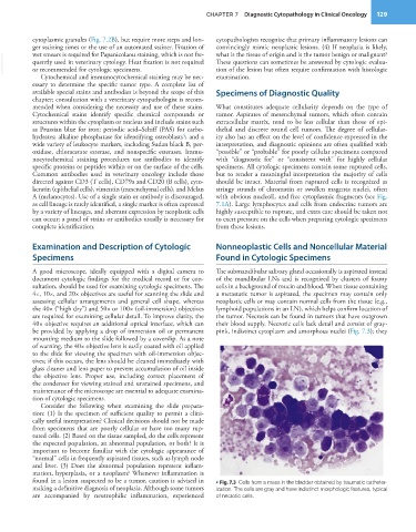
           be provided by applying a drop of immersion oil or permanent   pink, indistinct cytoplasm and amorphous nuclei (Fig. 7.3); they
           mounting medium to the slide followed by a coverslip. As a note
           of warning, the 40× objective lens is easily coated with oil applied
           to the slide for viewing the specimen with oil-immersion objec-
           tives; if this occurs, the lens should be cleaned immediately with
           glass cleaner and lens paper to prevent accumulation of oil inside
           the objective lens. Proper use, including correct placement of
           the condenser for viewing stained and unstained specimens, and
           maintenance of the microscope are essential to adequate examina-
           tion of cytologic specimens.
             Consider the following when examining the slide prepara-
           tion: (1) Is the specimen of sufficient quality to permit a clini-
           cally useful interpretation? Clinical decisions should not be made
           from specimens that are poorly cellular or have too many rup-
           tured cells. (2) Based on the tissue sampled, do the cells represent
           the expected population, an abnormal population, or both? It is
           important to become familiar with the cytologic appearance of
           “normal” cells in frequently aspirated tissues, such as lymph node
           and  liver. (3) Does  the  abnormal population  represent inflam-
           mation, hyperplasia, or a neoplasm? Whenever inflammation is
           found in a lesion suspected to be a tumor, caution is advised in   • Fig. 7.3  Cells from a mass in the bladder obtained by traumatic catheter-
           making a definitive diagnosis of neoplasia. Although some tumors   ization. The cells are gray and have indistinct morphologic features, typical
           are accompanied by neutrophilic inflammation, experienced   of necrotic cells.

