
Page 156 - Withrow and MacEwen's Small Animal Clinical Oncology, 6th Edition
P. 156
CHAPTER 7 Diagnostic Cytopathology in Clinical Oncology 135
pleomorphic TCCs must be differentiated from hyperplastic Renal Carcinomas
transitional epithelium that occurs secondary to inflammatory Renal carcinomas have few defining cytologic characteristics.
Variably pleomorphic cuboidal epithelial cells may be arranged
processes in the bladder; this can be challenging because inflam-
VetBooks.ir mation sometimes accompanies TCCs. Transitional cell polyps are in loose sheets, clusters, tubules, and acini. The cells have moder-
sampled infrequently and typically consist of sheets of epithelial
ate-to-high N:C ratios and may contain a few discrete cytoplas-
cells with a uniform or mildly pleomorphic appearance. mic vacuoles. Nuclei are generally round and centrally or basally
located, with variably distinct nucleoli. Cytologically, renal carci-
Tumors of Organs
nomas may be mistaken for neuroendocrine tumors.
Hepatocellular Tumors
In the liver, primary tumors may arise from hepatocytes or from Pulmonary Carcinomas or Adenocarcinomas
biliary epithelium. Hepatic carcinoids may be considered as pri- Pulmonary carcinomas or adenocarcinomas may occur in ani-
marily hepatic in origin (see Neuroendocrine Tumors). Hepa- mals with respiratory signs or may be found incidentally when
tocellular tumors include benign adenomas, or hepatomas, and thoracic radiographs are taken for another reason. Cats with pri-
carcinomas. Unfortunately, hepatic nodules and masses, whether mary pulmonary tumors may be presented for lameness result-
areas of hyperplasia, regeneration, benign tumors, or malignant ing from metastasis to the digits. Primary lung tumors are often
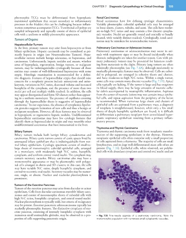
tumors, may be indistinguishable cytologically because all these minimally pleomorphic (see Fig. 7.4A), although moderately to
entities may consist of well-differentiated hepatocytes with some markedly pleomorphic features may be observed. Cells are cuboi-
atypia. Histologic examination is recommended for a defini- dal to polygonal, are arranged in cohesive sheets and clusters,
tive diagnosis. Features of hepatocellular atypia that should raise and have moderate-to-high N:C ratios. Within a single tumor,
concern for a neoplastic process include anisocytosis and aniso- some cells may contain many discrete vacuoles (Fig. 7.15). Apical
karyosis, variations in N:C ratios, decreased volume and increased cilia typically are lacking. If the tumor is large and has outgrown
basophilia of the cytoplasm, and the presence of more than two its blood supply, there may be large amounts of necrotic cellu-
nuclei per cell and multiple visible nucleoli. In addition, the cells lar debris accompanied by neutrophilic inflammation. Aspirates
may appear disorganized and form 3D clusters rather than appear- from the center of necrotic lesions may not contain intact epithe-
ing in a uniform monolayer. The presence of capillaries coursing lial cells, and repeat aspiration from the periphery of the lesion
through the hepatocellular sheets is suggestive of hepatocellular is recommended. When numerous large sheets and clusters of
7
carcinoma. In our experience, the absence of cytoplasmic lipofus- epithelial cells are aspirated from a pulmonary mass, a diagnosis
cin granules suggests formation of new cells and thus a benign or of neoplasia is straightforward; however, when only a few small
malignant neoplasm. However, all these features may be observed sheets of deeply basophilic epithelium are found, it is difficult
in hyperplastic or regenerative hepatic nodules. Undifferentiated to differentiate a pulmonary neoplasm from consolidated hyper-
hepatocellular carcinomas may have few cytologic features that plastic respiratory epithelium resulting from a primary inflam-
identify them as hepatocellular in origin and may resemble other matory process.
undifferentiated carcinomas that have metastasized to the liver.
Thymoma and Thymic Carcinoma
Biliary Tumors Thymoma and thymic carcinoma result from neoplastic transfor-
Biliary tumors include both benign biliary cystadenomas and mation of the supporting epithelium in the thymus. However,
carcinomas. Biliary cystic tumors consist of cystic spaces lined by neoplastic epithelial cells often comprise only a small proportion
attenuated biliary epithelium that is indistinguishable from nor- of cells aspirated from a thymoma. The majority of cells are small
mal biliary epithelium. Cytologic specimens consist of small-to- lymphocytes, and in dogs well-differentiated mast cells often are
large sheets of monomorphic cuboidal epithelial cells, arranged present (Fig. 7.16). Epithelial cells, when observed, are polyhe-
in a monolayer, with moderately high N:C ratios, basophilic dral cells with abundant cytoplasm and central oval nuclei and are
cytoplasm, and uniform central round nuclei. The cytoplasm may
contain secretory vacuoles. Biliary carcinomas also may have a
monomorphic appearance or may be pleomorphic with polygo-
nal cells arranged in sheets and 3D clusters; in this case, the cells
may have variable N:C ratios, deeply basophilic cytoplasm, and
central-to-eccentric oval nuclei. Secretory vacuoles may be numer-
ous, single, or absent. Nuclear and nucleolar pleomorphism is
prominent.
Tumors of the Exocrine Pancreas
Tumors of the exocrine pancreas may arise from ductular or acinar
epithelium. Cells from ductular carcinomas resemble biliary carci-
nomas and consist of monomorphic sheets of cuboidal cells with
high N:C ratios, basophilic cytoplasm, and central round nuclei.
Nuclear pleomorphism is typically mild, but criteria of malignancy
may be present. Exocrine pancreatic adenocarcinoma typically has
markedly pleomorphic features. The distinctive cytoplasm of exo-
crine pancreas, consisting of intensely basophilic cytoplasm with
numerous small eosinophilic globules, may be observed in a pro- • Fig. 7.15 Fine-needle aspirate of a pulmonary carcinoma. Note the
portion of cells supporting pancreatic origin. monomorphic population with numerous small cytoplasmic vacuoles.