Page 159 - Withrow and MacEwen's Small Animal Clinical Oncology, 6th Edition
P. 159
138   PART II    Diagnostic Procedures for the Cancer Patient


  VetBooks.ir


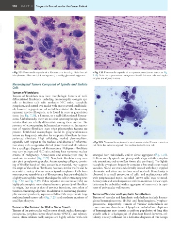
         • Fig. 7.21  Fine-needle aspirate of a fibrosarcoma in a dog. Note the cel-  • Fig. 7.22  Fine-needle aspirate of a myxosarcoma (same tumor as Fig.
         lular pleomorphism and pink background, possibly glycosaminoglycans.  7.19). Note the myxomatous background in which tumor cells and eryth-
                                                               rocytes are aligned in rows.

         Mesenchymal Tumors Composed of Spindle and Stellate
         Cells
         Tumors of Fibroblasts
         Tumors  of  fibroblasts  may  have morphologic  features  of  well-
         differentiated fibroblasts, including monomorphic elongate spi-
         ndle or fusiform cells with moderate N:C ratios, basophilic
         cytoplasm, and central oval nuclei with one to several small nucle-
         oli; however, a population of well-differentiated fibroblasts may
         represent reactive fibroplasia, as is found in scars or granulation
         tissue (see Fig. 7.20), a fibroma, or a well-differentiated fibrosar-
         coma. Unfortunately, there are no clear cytomorphologic charac-
         teristics that can reliably differentiate among these entities. The
         presence of accompanying inflammation warrants an interpreta-
         tion of reactive fibroblasts even when pleomorphic features are
         present. Epithelioid macrophages found in pyogranulomatous
         lesions are frequently mistaken for neoplastic fibroblasts by inex-
         perienced clinicians. High cellularity, marked pleomorphism,
         especially with respect to the nucleus, and absence of inflamma-  • Fig. 7.23  Fine-needle aspirate of a vaccine-associated fibrosarcoma in a
         tion along with a supportive clinical picture lend credible evidence   cat. Note the extreme atypia in the multinucleated tumor cell.
         for a cytologic diagnosis of fibrosarcoma. Malignant fibroblasts
         may vary in shape and N:C ratio and may have numerous nuclear
         criteria  of malignancy. Anisocytosis and anisokaryosis may be   arranged both individually and in dense aggregates (Fig. 7.24).
         moderate to marked (Fig. 7.21). Neoplastic fibroblasts may con-  Cells are usually spindle and plump with wispy veil-like cytoplas-
         tain pink cytoplasmic granules. Accompanying collagen, consist-  mic extensions; oval-to-stellate forms also are found. The lightly
         ing of fibrillar bands of pink extracellular material, may support   basophilic cytoplasm frequently contains a few small clear round
         the origin of the cells as fibroblasts; however, similar matrix can be   vacuoles. Nuclei are oval and centrally located with finely stippled
         seen with a variety of other mesenchymal neoplasms. Cells from   chromatin and often one to three small nucleoli. Binuclearity is
         myxosarcoma resemble cells of fibrosarcoma, but are embedded in   observed in a small proportion of cells, and multinucleate cells
         a lightly eosinophilic matrix that aligns the cells in streaming rows   with peripheralized nuclei, so-called “crown cells,” may be noted.
         (Fig. 7.22; see Fig. 7.19). Feline injection site sarcomas (ISSs) are   Anisocytosis and anisokaryosis are mild to moderate. Linear capil-
         highly pleomorphic mesenchymal tumors, primarily of fibroblas-  laries may be embedded within aggregates of tumor cells in aspi-
         tic origin, that occur at sites of previous injections, most often of   rates of perivascular wall tumors.
         vaccines containing adjuvant. In addition to containing pleomor-
         phic mesenchymal cells, aspirates of feline ISSs may contain large   Tumors of Vascular and Lymphatic Endothelium
         multinucleated tumor cells (Fig. 7.23) and moderate numbers of   Tumors of vascular and lymphatic endothelium include heman-
         small lymphocytes.                                    gioma/hemangiosarcoma  (HSA)  and  lymphangioma/lymphan-
                                                               giosarcoma,  respectively.  Tumors  of  vascular  endothelium  are
         Tumors of the Perivascular Wall or Nerve Sheath       more common than those of lymphatic endothelium. Aspirates
         Tumors of the perivascular wall or nerve sheath, such as hemangio-  of hemangioma may contain a uniform population of long thin
         pericytoma, peripheral nerve sheath tumor (PNST), and schwan-  spindle cells in a background of abundant blood; however, cel-
         noma, often exfoliate well; samples are highly cellular with cells   lularity is rarely sufficient for a definitive diagnosis of this benign

