Page 164 - Withrow and MacEwen's Small Animal Clinical Oncology, 6th Edition
P. 164
CHAPTER 7  Diagnostic Cytopathology in Clinical Oncology  143


  VetBooks.ir


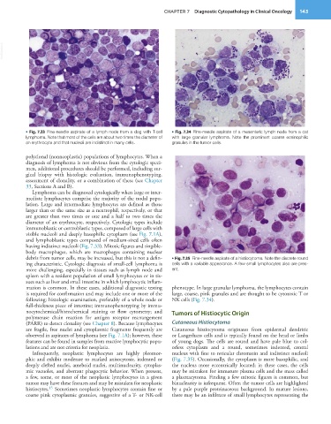
           • Fig. 7.33  Fine-needle aspirate of a lymph node from a dog with T-cell   • Fig. 7.34  Fine-needle aspirate of a mesenteric lymph node from a cat
           lymphoma. Note that most of the cells are about two times the diameter of   with large granular lymphoma. Note the prominent coarse eosinophilic
           an erythrocyte and that nucleoli are indistinct in many cells.  granules in the tumor cells.

           polyclonal (nonneoplastic) populations of lymphocytes. When a
           diagnosis of lymphoma is not obvious from the cytologic speci-
           men, additional procedures should be performed, including sur-
           gical  biopsy  with  histologic  evaluation,  immunophenotyping,
           assessment of clonality, or a combination of these (see Chapter
           33, Sections A and B).
             Lymphoma can be diagnosed cytologically when large or inter-
           mediate lymphocytes comprise the majority of the nodal popu-
           lation. Large and intermediate lymphocytes are defined as those
           larger than or the same size as a neutrophil, respectively, or that
           are greater than two times or one and a half to two times the
           diameter of an erythrocyte, respectively. Cytologic types include
           immunoblastic or centroblastic types, composed of large cells with
           visible nucleoli and deeply basophilic cytoplasm (see Fig. 7.1A),
           and lymphoblastic types composed of medium-sized cells often
           having indistinct nucleoli (Fig. 7.33). Mitotic figures and tingible-
           body macrophages, which are macrophages containing nuclear
           debris from tumor cells, may be increased, but this is not a defin-  • Fig. 7.35  Fine-needle aspirate of a histiocytoma. Note the discrete round
           ing characteristic. Cytologic diagnosis of small-cell lymphoma is   cells with a variable appearance. A few small lymphocytes also are pres-
           more challenging, especially in tissues such as lymph node and   ent.
           spleen with a resident population of small lymphocytes or in tis-
           sues such as liver and small intestine in which lymphocytic inflam-
           mation is common. In these cases, additional diagnostic testing   phenotype. In large granular lymphoma, the lymphocytes contain
           is required for confirmation and may include one or more of the   large, coarse, pink granules and are thought to be cytotoxic T or
           following: histologic examination, preferably of a whole node or   NK cells (Fig. 7.34).
           full-thickness piece of intestine; immunophenotyping by immu-
           nocytochemical/histochemical  staining  or  flow  cytometry;  and   Tumors of Histiocytic Origin
           polymerase chain reaction for antigen receptor rearrangement
           (PARR) to detect clonality (see Chapter 8). Because lymphocytes   Cutaneous Histiocytoma
           are fragile, free nuclei and cytoplasmic fragments frequently are   Cutaneous  histiocytoma  originates  from  epidermal  dendritic
           observed in aspirates of lymphoma (see Fig. 7.1A); however, these   or Langerhans cells and is typically found on the head or limbs
           features can be found in samples from reactive lymphocytic popu-  of young dogs. The cells are round and have pale blue to col-
           lations and are not criteria for neoplasia.           orless cytoplasm and a round, sometimes indented, central
             Infrequently,  neoplastic  lymphocytes  are  highly  pleomor-  nucleus with fine to reticular chromatin and indistinct nucleoli
           phic and exhibit moderate to marked anisocytosis, indented or   (Fig. 7.35). Occasionally, the cytoplasm is more basophilic, and
           deeply clefted nuclei, ameboid nuclei, multinuclearity, cytoplas-  the nucleus more eccentrically located; in these cases, the cells
           mic vacuoles, and aberrant phagocytic behavior. When present,   may be mistaken for immature plasma cells and the mass called
           a few, some, or most of the neoplastic lymphocytes in a given   a plasmacytoma. Finding a few mitotic figures is common, but
           tumor may have these features and may be mistaken for neoplastic   binuclearity is infrequent. Often the tumor cells are highlighted
                   15
           histiocytes.  Sometimes neoplastic lymphocytes contain fine or   by a pale purple proteinaceous background. In mature lesions,
           coarse pink cytoplasmic granules, suggestive of a T- or NK-cell   there may be an infiltrate of small lymphocytes representing the

