Page 162 - Withrow and MacEwen's Small Animal Clinical Oncology, 6th Edition
P. 162
CHAPTER 7  Diagnostic Cytopathology in Clinical Oncology  141


  VetBooks.ir


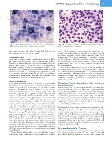
           • Fig. 7.30  Fine-needle aspirate of a melanoma in a dog. Note the fine   • Fig. 7.31  Fine-needle aspirate of a melanoma in which melanin granules
           melanin granules in the tumor cells and in the background.  are not visible (amelanotic melanoma).

           granules are common, and nuclei are round with finely stippled   pigmented melanomas contain melanophages, which are mac-
           chromatin and variably prominent nucleoli.            rophages containing  abundant melanin; in the authors’ experi-
                                                                 ence, this is especially true after surgical removal or biopsy of the
           Periarticular Tumors                                  primary mass. These melanophages may be mistaken for meta-
           Periarticular tumors are predominantly histiocytic sarcomas (HSs)   static cells, but they differ from neoplastic melanoblasts as mela-
           with other sarcomas (synovial myxoma, pleomorphic sarcoma,   nophages typically contain coarse collections of melanin within
           fibrosarcoma, and undifferentiated sarcoma) diagnosed less fre-  phagolysosomes rather than the fine granulation typically found
                 11
           quently.  Descriptions of synovial cell sarcoma histologically as   within melanoblasts.
           monophasic (spindle) or biphasic (epithelioid and spindle) may   Most dermal tumors of melanocytic origin are benign and
           encompass these different tumor types or a periarticular tumor of   are termed melanocytomas. They consist of polygonal or spindle
           undefined cell lineage. 12,13  Cytomorphologic appearances of peri-  cells containing black cytoplasmic granules. Several of the adnexal
           articular tumors correspond to the specific tumor type described   tumors may contain melanin pigment and must be differentiated
           elsewhere under mesenchymal tumors with round or spindle cells   from melanocytomas. These typically are of epithelial origin and
           or under tumors with frequent multinucleated cells.   should be distinguished by their cellular arrangement in cohesive
                                                                 sheets (see Fig. 7.9).
           Tumors of Melanocytes
           In malignant melanoma, the cells can adopt the appearance of   Mesenchymal Tumors Composed of Cells Arranged in
           epithelial (sheets of cohesive cells), mesenchymal (individual-  Dense Aggregates
           ized oval or spindle cells), or discrete round cell tumors, and all   Cells aspirated from some mesenchymal tumors, including rhab-
           three cytologic appearance may be evident in the same tumor.   domyosarcoma, perivascular wall tumor, PNST, amelanotic mela-
           Individual melanoblasts are round, oval, or spindle cells with   noma, and the epithelioid form of HSA, form dense aggregates
           moderately high N:C ratios, lightly basophilic cytoplasm, and   and clusters that are more characteristic of epithelial cells. Careful
           round or oval nuclei with fine chromatin and distinct nucleoli.   examination typically reveals some spindle cells with indistinct
           Criteria of malignancy consist primarily of anisokaryosis and   margins and spaces between the closely packed cells indicating
           nucleolar pleomorphism. Highly pigmented tumors do not   the lack of intercellular junctions. Slide preparation by imprinting
           present a diagnostic challenge, and fine black melanin gran-  and scraping also may yield clusters and sheets of mesenchymal
           ules may be so numerous that they obscure all cellular detail.   cells that mimic epithelial populations.
           Cells with varying degrees of pigmentation are typically found
           within the same tumor, and melanin granules may be sparse   Mesenchymal Tumors with Frequent Multinucleated Cells
           in some cells (Fig. 7.30). Amelanotic tumors present a greater   Although  any  neoplasm  can  have a  few  multinucleated  cells,
           diagnostic challenge. Usually, a faint scattering of fine gray-  multinuclearity is especially common in certain sarcomas. These
           black  melanin  granules  is  found  in  a  few  cells  to  support  a   include HSs, feline ISSs, pleomorphic sarcoma (also called malig-
           diagnosis, but cells may be completely devoid of pigmentation   nant fibrous histiocytosis or anaplastic sarcoma with giant cells),
           (Fig. 7.31). In these circumstances, moderately pleomorphic   rhabdomyosarcoma, and some plasmacytomas in which the mul-
           tumor cells aspirated from masses on the digits or in the oral   tinucleated cells are part of the tumor population. In OSA, mul-
           cavity should alert the clinician to the possibility of this highly   tinucleated osteoclasts are often present and are not part of the
           malignant tumor. Because melanocytes are of neuroectodermal   neoplastic population of cells.
           origin, the cells may express certain neural markers, such as
           S-100 and neuron-specific enolase, in addition to vimentin and   Discrete Round Cell Tumors
           often Melan A.
             An additional cytologic challenge is the identification of meta-  The majority of discrete round cell tumors are of hematopoi-
           static lesions within lymph nodes. Most lymph nodes draining   etic origin, including neoplasms of mast cells, plasma cells,

