Page 187 - Withrow and MacEwen's Small Animal Clinical Oncology, 6th Edition
P. 187
166   PART III    Therapeutic Modalities for the Cancer Patient


  VetBooks.ir


                           C


                              D
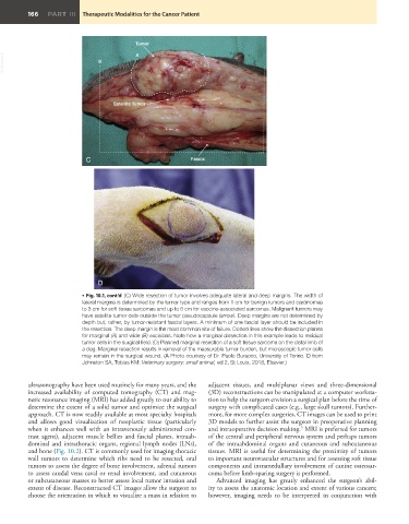
                          • Fig. 10.1, cont’d  (C) Wide resection of tumor involves adequate lateral and deep margins. The width of
                          lateral margins is determined by the tumor type and ranges from 1 cm for benign tumors and carcinomas
                          to 3 cm for soft tissue sarcomas and up to 5 cm for vaccine-associated sarcomas. Malignant tumors may
                          have satellite tumor cells outside the tumor pseudocapsule (arrow). Deep margins are not determined by
                          depth but, rather, by tumor-resistant fascial layers. A minimum of one fascial layer should be included in
                          the resection. The deep margin is the most common site of failure. Dotted lines show the dissection planes
                          for marginal (A) and wide (B) excisions. Note how a marginal dissection in this example leads to residual
                          tumor cells in the surgical field. (D) Planned marginal resection of a soft tissue sarcoma on the distal limb of
                          a dog. Marginal resection results in removal of the measurable tumor burden, but microscopic tumor cells
                          may remain in the surgical wound. (A Photo courtesy of Dr. Paolo Buracco, University of Torino. D from
                          Johnston SA, Tobias KM: Veterinary surgery: small animal, ed 2, St Louis, 2018, Elsevier.)


         ultrasonography have been used routinely for many years, and the   adjacent tissues, and multiplanar views and three-dimensional
         increased availability of computed tomography (CT) and mag-  (3D) reconstructions can be manipulated at a computer worksta-
         netic resonance imaging (MRI) has added greatly to our ability to   tion to help the surgeon envision a surgical plan before the time of
         determine the extent of a solid tumor and optimize the surgical   surgery with complicated cases (e.g., large skull tumors). Further-
         approach. CT is now readily available at most specialty hospitals   more, for more complex surgeries, CT images can be used to print
         and allows good visualization  of neoplastic tissue (particularly   3D models to further assist the surgeon in preoperative planning
                                                                                           5
         when it enhances well with an intravenously administered con-  and intraoperative decision making.  MRI is preferred for tumors
         trast agent), adjacent muscle bellies and fascial planes, intraab-  of the central and peripheral nervous system and perhaps tumors
         dominal and intrathoracic organs, regional lymph nodes (LNs),   of the intraabdominal organs and cutaneous and subcutaneous
         and bone (Fig. 10.2). CT is commonly used for imaging thoracic   tissues. MRI is useful for determining the proximity of tumors
         wall  tumors  to  determine  which  ribs  need  to  be  resected,  oral   to important neurovascular structures and for assessing soft tissue
         tumors to assess the degree of bone involvement, adrenal tumors   components and intramedullary involvement of canine osteosar-
         to assess caudal vena caval or renal involvement, and cutaneous   coma before limb-sparing surgery is performed.
         or subcutaneous masses to better assess local tumor invasion and   Advanced imaging  has greatly enhanced the surgeon’s abil-
         extent of disease. Reconstructed CT images allow the surgeon to   ity to assess the anatomic location and extent of various cancers;
         choose the orientation in which to visualize a mass in relation to   however, imaging needs to be interpreted in conjunction with

