Page 188 - Withrow and MacEwen's Small Animal Clinical Oncology, 6th Edition
P. 188
CHAPTER 10  Surgical Oncology  167


                                                                  TABLE 10.1     Classification and Resection of Wound
                                                                             Margins
  VetBooks.ir                                                      Type       Plane of Dissection    Result
                                                                   Intracapsular
                                                                              Tumor removed piecemeal or
                                                                                                     Residual macro-
                                                                                curetted, “debulking”  scopic disease
                                                                   Marginal   Tumor removed on or adjacent   Usually leaves
                                                                                to the tumor pseudocapsule,   microscopic
                                                                                “shelled out”          disease
                                                                   Wide       Tumor removed with margins of   Possible skip
                                                                                normal tissue lateral and deep   lesions
                                                                                to the tumor; tumor capsule is
                                                                                not compromised
                                                                   Radical    Tumor removed with an entire   No local residual
                                                                                compartment or structure    cancer
                                                                                (e.g., amputation)
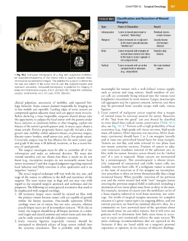
           •  Fig. 10.2  Computed tomography of a dog with suspected multilobu-
           lar osteochondrosarcoma of the inferior orbit is used to provide three-
           dimensional reconstructed images. This enables the surgeon to determine
           the size and extent of the tumor and to plan the surgical margins and
           approach accurately. Computed tomography is preferred for imaging of   meaningful for tumors with a well-defined venous supply,
           bone and intrathoracic masses. (From Johnston SA, Tobias KM: Veterinary
           surgery: small animal, ed 2, St Louis, 2018, Elsevier.)  such as splenic and lung tumors. Small numbers of can-
                                                                   cer cells are constantly being released into the venous (and
                                                                   lymphatic) circulation by most tumors. Larger, macroscopic
           clinical palpation, assessment of mobility, and expected bio-  cell aggregates may be a greater concern, however, and these
           logic behavior. Some cancers deemed inoperable by imaging are   may be prevented from vascular escape with early venous
           in fact mobile and operable. Leading edges of some cancers are   ligation.
           compressed against adjacent tissue and can appear more invasive.    3.   Local control of malignant cancer requires that a margin
           Before declaring a mass inoperable, surgeons should always take   of normal tissue be removed around the tumor. Resection
           the opportunity to palpate the local tumor with the patient under   of the “bad from the good” can and should be classified
           heavy sedation or anesthesia before or after imaging, explore the   in more detail than radical versus conservative (Table 10.1;
                                                                                  8
           history of the tumor’s growth pattern and, in many cases, obtain a   also, see Fig. 10.1).  Tumors with a high probability of local
           tissue sample. Positive prognostic factors typically include a slow   recurrence (e.g., high-grade soft tissue sarcoma, high-grade
           growth rate, mobility within adjacent tissues, no previous surgery,   mast cell tumors, feline injection-site sarcomas, feline mam-
           discrete tumor borders, small tumor size, and a low-grade nature.   mary carcinoma) should have 2 to 3 cm margins laterally
           Conversely, surgery may be less effective for the same tumor type   and at least one uninvolved fascial layer for deep margins.
           and grade if the mass is ill defined, recurrent, or has a recent his-  Tumors are not flat, and wide removal in one plane does
           tory of rapid growth.                                   not ensure complete excision. Fixation of cancer to adja-
             The surgical oncologist must be able to assimilate all of the   cent structures mandates removal of the adherent area en
           information and make an informed decision.  We must also   bloc with the tumor. Invasive cancer should not be “shelled
           remind ourselves and our clients that there is much we do not   out” if a cure is expected. Many cancers are surrounded
           know (e.g., incomplete margins do not necessarily ensure local   by a pseudocapsule. This pseudocapsule is almost invari-
           tumor recurrence ) and that surgical judgment regarding expected   ably composed of compressed and viable tumor cells, not
                        6
           local behavior and likely resection is often qualitative and is an   healthy, reactive host cells. If a malignant tumor is entered
           imperfect “science.”                                    at the time of resection or if the margins are incomplete,
             The actual surgical technique will vary with the site, size, and   that procedure is often no better therapeutically than a large
           stage of the tumor, in addition to the skill and experience of the   incisional biopsy. When possible, resection of the previous
           surgeon. The same tumor type in dogs and cats may vary with   scar and the entire wound bed with “new” margins (never
           regard to the required surgical approach and technique and the   entering the previous surgical field) is indicated, including a
           prognosis. The following are some general statements that need to   minimum of one tissue plane away from or deep to the mass.
           be emphasized with surgical oncology.                   For example, invasion of cancer into the medullary cavity of
            1.   All incisional biopsy  tracts  should be excised  en bloc with   a bone requires subtotal or total bone resection, not curet-
             the primary tumor because tumor cells are capable of growth   tage. The width of surgical margins necessary for complete
             within the biopsy incisions. Fine-needle aspiration (FNA)   excision of a given tumor type is an ongoing debate, and our
             cytology tracts are of minor, but not zero, concern, whereas   current practices are based on minimal objective data. As a
             punch biopsy tracts are of intermediate concern.  With this in   community we have answered most of the questions about
                                                  7
             mind, the surgeon should keep all biopsy incisions to a mini-  how much tissue we can safely remove, but it will serve our
             mal length and should position and orient them such that they   patients well to determine how little extra tissue is neces-
             can be easily removed with the definitive resection.  sary to excise and consistently achieve the same success. We
            2.   Early vascular ligation (especially venous) should be   must challenge recommendations that are reported in the
             attempted to diminish release of large tumor emboli into   literature  if  they  are  based  solely  on  a  surgeon’s  personal
             the systemic circulation. This is probably only clinically   experience or opinion, in the absence of objective findings.

